. 2023 May 29;35(6):2391-2412.
doi: 10.1093/plcell/koad064.

Phosphorylation and ubiquitination of OsWRKY31 are integral to OsMKK10-2-mediated defense responses in rice

Affiliations

Phosphorylation and ubiquitination of OsWRKY31 are integral to OsMKK10-2-mediated defense responses in rice

Shuai Wang et al. Plant Cell. .

Abstract

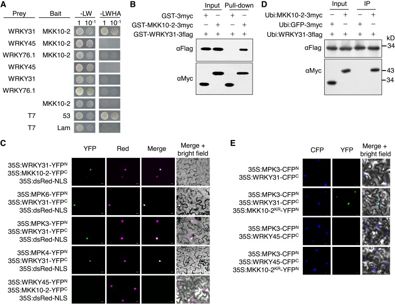
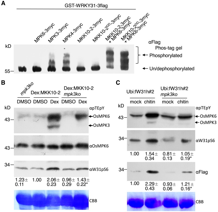
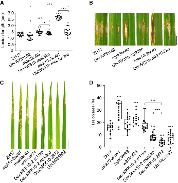
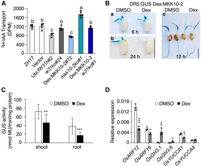
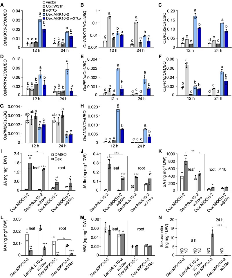
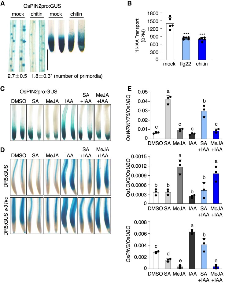
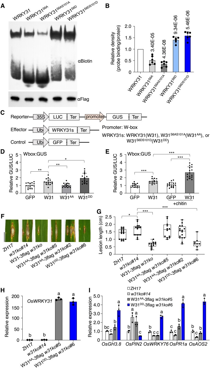
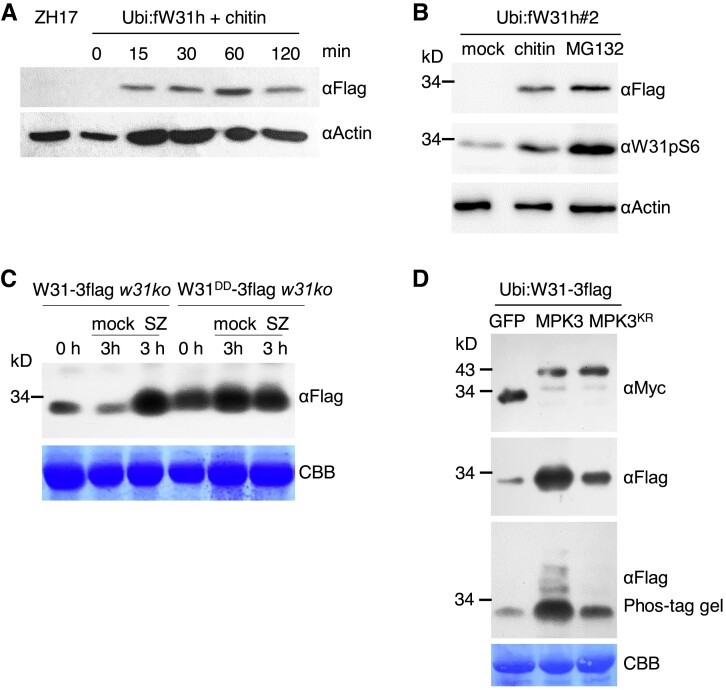
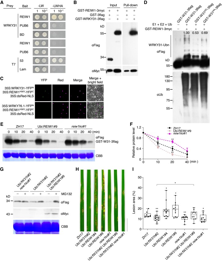
Mitogen-activated protein kinase (MPK) cascades play vital roles in plant innate immunity, growth, and development. Here, we report that the rice (Oryza sativa) transcription factor gene OsWRKY31 is a key component in a MPK signaling pathway involved in plant disease resistance in rice. We found that the activation of OsMKK10-2 enhances resistance against the rice blast pathogen Magnaporthe oryzae and suppresses growth through an increase in jasmonic acid and salicylic acid accumulation and a decrease of indole-3-acetic acid levels. Knockout of OsWRKY31 compromises the defense responses mediated by OsMKK10-2. OsMKK10-2 and OsWRKY31 physically interact, and OsWRKY31 is phosphorylated by OsMPK3, OsMPK4, and OsMPK6. Phosphomimetic OsWRKY31 has elevated DNA-binding activity and confers enhanced resistance to M. oryzae. In addition, OsWRKY31 stability is regulated by phosphorylation and ubiquitination via RING-finger E3 ubiquitin ligases interacting with WRKY 1 (OsREIW1). Taken together, our findings indicate that modification of OsWRKY31 by phosphorylation and ubiquitination functions in the OsMKK10-2-mediated defense signaling pathway.


Conflict of interest statement

Conflict of interest statement. The authors declare no conflict of interests.

Figures

Figure 1.
Figure 1.
OsMKK10-2 and OsMPKs interact with OsWRKY31. A) OsWRKY31, OsWRKY45, OsWRKY76.1, and OsMKK10-2 were fused to the Gal4 activation domain (prey) or DNA-binding domain (bait). The resulted plasmids in appropriate combinations were introduced into yeast cells and then incubated in synthetic dropout medium without Leu and Trp (-LW) or lacking Leu, Trp, His, and adenine (-LWHA) and photographed 3 d after plating. Yeast cells containing AD-T7 plus BD-53, and AD-T7 plus BD-Lam plasmids were used as the positive and negative controls, respectively. B) OsMKK10-2 and OsWRKY31 were sandwiched with GST and 3×Myc (GST-MKK10-2-3myc) or GST and 3×Flag (GST-WRKY31-3flag) tags. Purified protein (each about 1 μg) was combined accordingly and incubated at 4 °C for 2 h in immunoprecipitation buffer. The protein mixture was precipitated with anti-c-Myc agarose affinity gels, separated on 10% SDS-PAGE gels, and detected by immunoblots with αMyc and αFlag antibodies. C) OsWRKY31, OsWRKY45, OsMKK10-2, OsMPK3, OsMPK4, and OsMPK6 were fused in frame with the YFP N-terminal region (YFPN) or the YFP C-terminal region (YFPC). The Agrobacteria with combined plasmids were infiltrated into the leaves of N. benthamiana. Confocal images were taken at 2 d after the infiltrations. Red fluorescence signals indicative of the nuclei were from co-infiltrated 35S:dsRed-NLS (NLS: nuclear localization signal). D) Plasmids of Ubi:MKK10-2-3myc, Ubi:WRKY31-3flag, and Ubi:GFP-3flag control were introduced separately in A. tumefaciens and co-expressed in appropriate combinations in leaves of N. benthamiana. Total proteins were extracted and immunoprecipitated with anti-c-Flag affinity gels. The precipitates were subjected for immunoblots with αMyc and αFlag antibodies. E) OsWRKY31 or OsWRKY45 and OsMPK3 were fused with CFPC and CFPN, respectively. OsMKK10-2KR, a kinase-dead mutant with Lys81 to Arg81 substitution, was fused with YFPN. Images photographed at 2 d after the infiltrations are CFP (left), YFP (middle), and merged with bright field (right). Bar = 10 μm.
Figure 2.
Figure 2.
OsMKK10-2 activates phosphorylation of OsMPKs and OsWRKY31. A) The recombinant proteins were expressed with GST plus 3×Myc or 3×Flag tag. GST-WRKY31-3flag with appropriate combination of kinase(s) were subjected to phosphorylation at 30 °C for 2 h. Proteins were separated on Phos-tag gels and blotted with αFlag antibody. The retarded bands indicated the phosphorylated GST-WRKY31-3flag. OsMKK10-2DD: mutations of Arg203 and Ser209 to Asp for constitutive activation. B) Seven-day-old seedlings cultured hydroponically were treated with 10 µM dexamethasone (Dex) or DMSO as the mock. Proteins extracted were separated on 10% SDS-PAGE gels and blotted with αpTEpY, αOsMPK6, or αW31pS6 (generated for detection of OsWRKY31 S6 phosphorylation) antibody. Representative data from four independent experiments. C) Ubi:fW31h#2 mpk3ko and Ubi:fW31h#2 seedlings were grown hydroponically for 7 d, and then treated with 200 µg mL−1 chitin. The proteins extracted were analyzed by immunoblots with αpTEpY, αW31pS6, and αFlag. CBB, Coomassie brilliant blue staining for loading control. Representative data from three independent experiments. Relative intensity (numbers below the gels) of bands was analyzed using ImageJ software. The values below indicate the relative gray density of three experiments. The value marked with * indicates statistically significant difference between Dex treatment B) and chitin C), analyzed by the Student's t-test (*, P < 0.05). Dex:MKK10-2 for Dex:MKK10-2-myc; mpk3ko for OsMPK3 knockout; Ubi:fW31h for Ubi:Flag-WRKY31-6×His.
Figure 3.
Figure 3.
OsWRKY31 and OsMPK3 are involved in OsMKK10-2 activated disease resistance. Lesion length A) and disease symptoms B) of the transgenic and wild-type ZH17 plants after infiltration with M. oryzae SZ strain. Plants of tillering stage (about 3-mo-old) in paddy field were injected with SZ spores (1 × 10⁵ conidia mL−1) in year 2020. The segments of leaves were photographed and quantified at 9 d after the infiltrations. The median was the crossline in each boxplot showing the lesion length distributions (n ≥ 9). Disease symptoms C) and lesion areas D) of the transgenic and ZH17 plants infected with SZ strain by spraying. Eighteen-day-old plants were inoculated with SZ spores (5 × 10⁵ conidia mL−1) by foliar spraying; the Dex-inducible lines were treated with 20 µM Dex for 12 h before the pathogen inoculation and the other plants were received with DMSO as the mock. Evaluation of disease severity and photography taken were conducted at 7 d after the inoculation. Ubi:fW31h mkk10-2ko for Ubi:fW31h and mkk10-2ko genetic crossing progeny; other plants described in Fig. 2. The median was the crossline in each boxplot showing the lesion area distributions (n = 16). Significance was evaluated by comparing with ZH17 or each other as bracketed using the Student's t-test (*, P < 0.05; **, P < 0.01; ***, P < 0.001). Bar = 1 cm.
Figure 4.
Figure 4.
Activation OsMKK10-2 suppresses auxin transport and homeostasis. A) Polar auxin transport (PAT) assay of coleoptiles. Seven-day-old seedlings growing in the dark were treated with 10 µM Dex for 12 h and then segmented for PAT assay. Values shown are means ± SD of five independent replicates. Values marked with different letters indicate statistically significant differences as analyzed by Duncan's multiple range test, α = 0.05. GUS staining B) and activity C) of DR5:GUS Dex:MKK10-2 (genetic crossing of DR5:GUS and Dex:MKK10-2) plants. Seedlings of 2 d after germination were treated with 10 µM Dex or DMSO for 6 (a) and 24 h (b). DR5:GUS Dex:MKK10-2 seedlings of 5 d old were treated with DMSO solvent or Dex for 12 h for GUS staining (c) and activity determination (n = 14) C). DR5:GUS for auxin responsive elements (DR5) controlled GUS reporter gene; Bar = 1 cm. D) Expression of auxin related genes. Dex:MKK10-2 seedlings of 12 d old were treated with 10 µM Dex for 6 h and DMSO as the mock, and the leaves were sampled for total RNA isolation. Gene expression was determined by RT-qPCR using OsUBQ as the reference gene. Results from representative experiments are shown. Experiments were repeated three times with similar results. Values are means ± SD with significant difference compared with DMSO mock using Student's t-test (*, P < 0.05; **, P < 0.01; ***, P < 0.001).
Figure 5.
Figure 5.
OsWRKY31 is required for OsMKK10-2-activated immune response. The transgenic and control plants were hydroponically cultured for 10 d, treated with 10 µM Dex, and sampled at designated time points for RNA and chemical isolations. Transcript level of each gene A to H) was determined by RT-qPCR using OsUBQ as the internal reference. Values represent means ± SD of three indepent treatments, each including three seedling leaves. Values marked with different letters indicate statistically significant differences as analyzed by the SPSS software (Duncan's multiple range test, α = 0.05). I to N) Contents of compounds were determined by liquid chromatography–tandem mass spectrometry using D6ABA as the internal standard. I to M) sampled at 6 h after the Dex treatment. N) Leaf samples at 6 and 24 h after the Dex treatment; ND, no detection; root × 10, indicating 10 times upscale the value in root. Data are means ± SD of three or four biological repeats. Significance test was evaluated using the Student's t-test by comparing with DMSO treatment or each other as bracketed (*, P < 0.05; **, P < 0.01; ***, P < 0.001).
Figure 6.
Figure 6.
OsMKK10-2 and OsWRKY31 are involved in PAMP regulation of defense and auxin pathways. A) Changes of OsPIN2pro:GUS expression pattern and primordia formation by chitin treatment. Five-day-old OsPIN2pro:GUS seedlings were treated with 200 µg mL−1 chitin for 12 h and stained at 37 °C for 4 h. Values are means ± SD of three biological replicates. B) Chitin and flg22 suppress auxin transport. Seven-day-old ZH17 seedlings grown in the dark were treated with 1 µM flg22 or 200 µg mL−1 chitin for 12 h, and then the coleoptiles were collected for PAT assay as described in Fig. 4. Error bars represent standard deviation of five replicate experiments. Significance test was evaluated using the Student's t-test (***, P < 0.001). Suppression of OsPIN2C) and DR5D) promoter activities by MeJA and SA. Five-day-old OsPIN2pro:GUS, DR5:GUS, and DR5:GUS w31ko seedlings were treated with MeJA (200 µM), SA (500 µM), or in combination with IAA (1 µM), and the mock received the same amount of DMSO at 28 °C for 12 h. Representative GUS staining images of OsPIN2pro:GUS primary roots stained at 37 °C for 4 h, and DR5:GUS and DR5:GUS w31ko staining at 37 °C for 1 h. Bar = 0.5 mm C and D). E) Expression of SA (OsWRKY76), JA (OsLOX2), and IAA (OsPIN2) marker genes in roots. Five-day-old DR5:GUS seedlings were treated with phytohormones as described in D). Gene expression was determined by RT-qPCR analysis using OsUBQ as the reference. Data are means ± SD of three independent treatments. Values marked with different letters indicate statistically significant differences as analyzed by the SPSS software (Duncan's multiple range test, α = 0.05).
Figure 7.
Figure 7.
Phosphomimetic OsWRKY31 enhances DNA-binding activity and disease resistance. A) Phosphomimetic (GST-WRKY31S6D-3flag and GST-WRKY31S6DS101D-3flag), phosphonull (GST-WRKY31S6A-3flag and GST-WRKY31S6AS101A-3flag), and GST-WRKY31-3flag protein were expressed in E. coli. and purified for electrophoretic mobility shift assay using BP22-f3 probe (biotin-labeled P22-f3 probe, Supplemental Fig. S14A). The mixture of protein (about 2 µg) with probe (0.03 μM) was separated on native PAGE gels and detected with αBiotin antibody. The presence of retarded band indicating the formation of protein–DNA complex. Protein loading was analyzed using αFlag antibody. B) The relative density is the retarded band in related to the loading protein and normalized with the wild-type OsWRKY31 protein. Values are means ± SD of six repeats. P-values are shown in the graph. C) Schematic diagrams of the reporter and effector constructs. The promoter was fused with GUS reporter gene. LUC gene driven by the cauliflower mosaic virus (CaMV) 35S was used as an internal control. The W-box DNA was put ahead of 35S minimal promoter with TATA-box to generate Wbox:GUS construct. D and E) Effector of OsWRKY31 (W31), phosphomimetic OsWRKY31S6DS101D (W31DD), phosphonull OsWRKY31S6AS101A (W31AA), or GFP control was co-infiltrated with the reporter of Wbox:GUS in the leaves of N. benthamiana. The treated tobacco plants were grown 25 °C for 3 d and sampled for GUS activity. In case of chitin treatment, the leaves were sprayed with chitin solution (200 µg mL−1) 5 h before the sample collection. Values are means ± SD of different leaf discs (n ≥ 14). Overexpression of phosphomimetic OsWRKY31DD elevated resistance to M. oryzae SZ strain. F and G) The phosphomimetic W31DD-3flag, the phosphonull W31AA-3flag, and W31-3flag genes (all controlled by ubiquitin promoter) were transformed into w31ko background. The hygromycin-resistant transgenics of T2 progenies and the ZH17 control were grown to the tillering stage in the paddy filed and inoculated with spores of M. oryzae SZ (1 × 10⁵ conidia mL−1) in year 2020. The leaf segments were photographed and quantified at 9 d after the infiltrations. The median was the crossline in each boxplot showing the lesion length distributions (n ≥ 8). Significance was evaluated by comparing with each other as bracketed using the Student's t-test (*, P < 0.05; **, P < 0.01; ***, P < 0.001). Bar = 1 cm. H and I) Gene expression. Leaves of 10-d-old seedlings cultured hydroponically were used for total RNA isolation. Gene expression was determined by RT-qPCR using OsUBQ as the reference gene. Values are means ± SD of three independent expereiments, each including three seedling leaves. Values marked with different letters indicate statistically significant differences as analyzed by the SPSS software (Duncan's multiple range test, α = 0.05).
Figure 8.
Figure 8.
Rapid induction of OsWRKY31 protein by chitin and M. oryzae. A and B) Induction of OsWRKY31 accumulation by chitin and proteasome inhibitor MG132. Leaves of 7-d-old Ubi:fW31h#2 were treated with 200 µg mL−1 chitin, 100 µM MG132 or DMSO as the mock for 1 h B) or with 200 µg mL−1 chitin at various time points A). The extracted proteins were analyzed by immunoblots with antibodies of αFlag, αW31pS6, and αActin as the loading control. C) Induction of OsWRKY31 protein by M. oryzae. Eighteen-day-old rice plants (Ubi:W31DD-3flag w31ko and Ubi:W31-3flag w31ko) growing in soil were inoculated with spores of M. oryzae SZ (5 × 10⁵ conidia mL−1) by spraying, and the leaves were sampled for protein analysis at 3 h after or before (0 h) the challenge. The proteins extracted were analyzed by immunoblots with αFlag antibody. Results from representative experiments are shown. Experiments were performed in three replicates. D) Increase OsWRKY31 accumulation by phosphorylation. Agrobacteria harboring Ubi:W31-3flag plasmid were co-infiltrated with agrobacteria containing 35S:MPK3-myc, 35S:MPK3KR-myc, or 35S:GFP-myc, into the leaves of N. benthamiana. Proteins were extracted from the leaves of 2 d after the infiltrations and analyzed by immunoblots with αMyc and αFlag antibodies. CBB, Coomassie brilliant blue staining for loading control. Representative data from three independent experiments.
Figure 9.
Figure 9.
OsREIW1 regulates OsWRKY31 stability. A to C) OsREIW1 interacts with OsWRKY31. A) OsREIW1 and PUB6 as a control were fused to the Gal4 DNA-binding domain (bait). The prey-OsWRKY31 was from Fig. 1. Yeast cells were incubated in SD medium without Leu and Trp (-LW) or lacking Leu, Trp, His, and adenine (-LWHA), and photographed 3 d after plating. Yeast cells containing AD-T7 plus BD-53 and AD-T7 plus BD-Lam plasmids were used as the positive and negative controls, respectively. B) Purified GST-REIW1-3myc and GST-WRKY31-3flag protein (each about 1 μg) were mixed and incubated at 4 °C for 2 h in immunoprecipitation buffer. The protein mixture was precipitated with anti-c-Myc agarose affinity gels, separated on 10% SDS-PAGE gels, and detected by immunoblots with αMyc and αFlag antibodies. C) Agrobacteria harboring 35S:REIW1H56Y-YFPC, 35S:WRKY31-YFPN or 35S:WRKY76.1-YFPN plasmid were combined accordingly and infiltrated into the leaves of N. benthamiana. Confocal images were taken at 2 d after the infiltrations. Red fluorescence signals indicative of the nuclei were from co-infiltrated 35S:dsRed-NLS (NLS: nuclear localization signal). Bar = 10 μm. D) Ubiquitination of OsWRKY31 by OsREIW1. OsWRKY31 recombinant protein GST-W31-3flag and its phosphomimetic GST-W31DD-3flag and phosphonull GST-W31AA-3flag were incubated with E1, E2, and ubiquitin in the presence of GST-REIW1-3myc. The reaction mixture (30 μL) contained 50 ng wheat ubiquitin-activating enzyme E1, 100 ng human ubiquitin-binding enzyme E2 (UBCH5b), 5 µg Arabidopsis ubiquitin (Ub), 500 ng GST-REIW1-3myc, and 2 μg OsWRKY31 or its mutant protein in buffer (50 mM Tris-HCl pH 7.5, 10 mM MgCl2, 2 mM DTT, and 5 mM ATP) and incubated at 30 °C for 1 h. The reaction was stopped by adding the 5× SDS loading buffer. Samples were separated by 10% SDS-PAGE gels and analyzed by immunoblots using αUb or αFlag antibody. Relative intensity (numbers in the gel) of ubiquitinated bands was analyzed using ImageJ software. E and F) Cell-free degradation of OsWRKY31. Total proteins were extracted from the leaves of Ubi:REIW1-3myc, reiw1ko, and ZH17 seedlings grown hydroponically for 1 wk. Protein extracts (about 500 μg) were incubated with GST-WRKY31-3flag (2 μg) for various time points at 25 °C. The protein samples were separated and analyzed by immunoblot with αFlag antibody. Representative data from three independent experiments. Quantification of treatments at 0 min was set as 1. Values are mean ± SD (n = 3). G) Segments of leaves from 7-d-old seedlings cultured hydroponically were treated with 100 µM MG132 or DMSO solvent for 1 h and then sampled for protein isolation. The protein mixture was separated on 10% SDS-PAGE gels and detected by immunoblots with αMyc and αFlag antibodies. Representative results from three independent experiments. CBB, Coomassie brilliant blue staining for loading control. H and I) Eighteen-day-old plants were inoculated with M. oryzae SZ spores (5 × 10⁵ conidia mL−1) by foliar spraying. Disease symptoms H) and lesion areas I) were conducted at 7 d after the inoculation. Representative data from three independent experiments. Significance was evaluated by comparing with ZH17 or each other as bracketed using Student's t-test (*, P < 0.05; **, P < 0.01). Bar = 2 cm.

Comment in

Similar articles

Cited by

References

    1. Achnine L, Blancaflor EB, Rasmussen S, Dixon RA. Colocalization of L-phenylalanine ammonia-lyase and cinnamate 4-hydroxylase for metabolic channeling in phenylpropanoid biosynthesis. Plant Cell. 2004:16(11): 3098–3109. 10.1105/tpc.104.024406 - DOI - PMC - PubMed
    1. Andreasson E, Ellis B. Convergence and specificity in the Arabidopsis MAPK nexus. Trends Plant Sci. 2010:15(2): 106–113. 10.1016/j.tplants.2009.12.001 - DOI - PubMed
    1. Cai Y, Chen X, Xie K, Xing Q, Wu Y, Li J, Du C, Sun Z, Guo Z. Dlf1, a WRKY transcription factor, is involved in the control of flowering time and plant height in rice. PLoS One. 2014:9(7): e102529. 10.1371/journal.pone.0102529 - DOI - PMC - PubMed
    1. Chen Q, Liu R, Wu Y, Wei S, Wang Q, Zheng Y, Xia R, Shang X, Yu F, Yang X, et al. . ERAD-related E2 and E3 enzymes modulate the drought response by regulating the stability of PIP2 aquaporins. Plant Cell. 2021:33(8): 2883–2898. 10.1093/plcell/koab141 - DOI - PMC - PubMed
    1. Chen X, Li C, Wang H, Guo Z. WRKY transcription factors: evolution, binding, and action. Phytopathol Res. 2019:1(1): 13. 10.1186/s42483-019-0022-x - DOI

Publication types

MeSH terms

Substances