. 2023 Apr 11;51(6):2931-2949.
doi: 10.1093/nar/gkad108.

Structural and functional insights into the activation of the dual incision activity of UvrC, a key player in bacterial NER

Affiliations

Structural and functional insights into the activation of the dual incision activity of UvrC, a key player in bacterial NER

Anna Seck et al. Nucleic Acids Res. .

Abstract

Bacterial nucleotide excision repair (NER), mediated by the UvrA, UvrB and UvrC proteins is a multistep, ATP-dependent process, that is responsible for the removal of a very wide range of chemically and structurally diverse DNA lesions. DNA damage removal is performed by UvrC, an enzyme possessing a dual endonuclease activity, capable of incising the DNA on either side of the damaged site to release a short single-stranded DNA fragment containing the lesion. Using biochemical and biophysical approaches, we have probed the oligomeric state, UvrB- and DNA-binding abilities and incision activities of wild-type and mutant constructs of UvrC from the radiation resistant bacterium, Deinococcus radiodurans. Moreover, by combining the power of new structure prediction algorithms and experimental crystallographic data, we have assembled the first model of a complete UvrC, revealing several unexpected structural motifs and in particular, a central inactive RNase H domain acting as a platform for the surrounding domains. In this configuration, UvrC is maintained in a 'closed' inactive state that needs to undergo a major rearrangement to adopt an 'open' active state capable of performing the dual incision reaction. Taken together, this study provides important insight into the mechanism of recruitment and activation of UvrC during NER.


Figures

Figure 1.
Figure 1.
Primary structure and domain organization of UvrC and characterization of its FeS cluster. (A) Schematic diagram of UvrC and its known domains and functional motifs. At the start of this study, little was known regarding the central domain (beige). The different constructs used in this study (UvrC, UvrC-Δ(HhH)2, UvrC-C, UvrC-N and UvrC-NEndo) are illustrated below. (B) Resonance Raman spectra of anaerobically purified DrUvrC (top trace), aerobically purified DrUvrC (middle trace) and the buffer (lower trace), measured with 406 nm excitation. Vibrational mode at 346 cm−1 is characteristic of [3Fe–4S]1+ cluster; the band at 420 cm−1, present also in the buffer, originates from glycerol. (C) Cyclic voltammograms of SAM-coated Au electrode before (dotted trace) and after immobilization of DrUvrC (solid trace). Measurements were performed in 50 mM Tris–HCl pH 7.5 and 100 mM NaCl at a scan rate of 50 mV s−1.
Figure 2.
Figure 2.
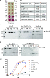
Analysis of the interaction between UvrB and UvrC. (A) Bacterial two hybrid analysis of the interaction between UvrB and UvrC. For each experiment, a bait and a prey corresponding to UvrB and UvrC proteins fused respectively to the T18 or T25 domains of bacterial adenylate cyclase, or the single T18 and T25 proteins alone, were co-transformed as indicated. Positive interactions were detected on MacConkey agar plates as red colored colonies. The cultures were spotted in duplicate. (B) Table presenting the size-exclusion chromatography (SEC) elution volumes of UvrB alone and in complex with different UvrC constructs. (C)-(D) SDS-PAGE analysis of the SEC fractions of the various UvrB/UvrC assemblies listed in (B). (C) UvrC constructs containing the N-terminal half of UvrC interact with UvrB. (D) UvrC-C and UvrC-NEndo do not interact with UvrB and elute as separate peaks. (E) DNA binding curves derived from fluorescence polarization (FP) measurements of 0–2 μM UvrB (green), UvrC (red), UvrC-Δ(HhH)2 (beige), UvrC-N (orange), UvrC-C (purple) or UvrB/UvrC (blue) binding to 2 nM 50 mer dsDNA containing a fluorescein-conjugated thymine in position 26 in the presence of ATP. Data points represent mean and standard deviation of three independent measurements and the data were fitted to a single specific binding model with Hill slope in GraphPad Prism 8. Derived binding constants are presented in Table 3.
Figure 3.
Figure 3.
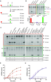
DrUvrABC incision assay. (A) Schematic diagram of the DrUvrABC incision assay relying on the use of D. radiodurans UvrA1, UvrB and UvrC proteins, ATP and Mg2+, and a dual labelled 50 mer dsDNA substrate containing a red fluorophore (ATTO633) on the 5′ end and a fluorescein conjugated thymine in position 26. (B) Representative TBE-polyacrylamide urea gel analysis of the DrUvrABC incision activity of UvrC point mutants. Reactions were performed for 1 hour at 37°C using 25 nM F26-seq1 substrate, 1 μM UvrA1, 0.5 μM UvrB and 2 μM of either UvrCWT, UvrCE72A or UvrCD391A in the presence of 2.5 mM Mg2+ and 2.5 mM ATP. The major bands observed by electrophoresis using either the red- (left gel) or green- (right gel) filter are indicated with arrows. These experiments were repeated three times and the quantitative assessments of these data are shown below the gels. Histograms present the mean and standard deviation of three independent reactions. Red bars correspond to the 18 mer fragment produced by 5′ cleavage, purple bars to the 30 mer fragment produced by 3′ cleavage, and green bars to the final 12 mer product resulting from dual incision. **** indicates a P value < 0.0001. (C) Representative TBE-polyacrylamide urea gel analysis of the DrUvrABC incision activity of single or combined UvrC truncation and point mutants. Reactions were performed for 1 hour at 37°C using 25 nM F26-seq1 substrate, 1 μM UvrA1, 0.5 μM UvrB and 2 μM of isolated UvrC domains either alone or in combination in the presence of 2.5 mM Mg2+ and 2.5 mM ATP. The major bands observed by electrophoresis using either the red- (top gel) or green- (lower gel) filter are indicated with arrows. These gels were repeated at least three times and the quantitative assessments of these data are presented in Supplementary Figure S9. The extent of 5′ and 3′ incision for each reaction is evaluated as – for no activity, + for low activity, ++ for slightly impaired activity and +++ for WT activity. (D) Kinetics of release of 12 mer product (full line) or 30 mer intermediate product (dashed line) by UvrC (black), UvrC-Δ(HhH)2 (red) and UvrC-Δ(HhH)2 + UvrC-C (orange). (E) Kinetics of release of 12 mer product (full line) or 30 mer intermediate product (dashed line) by UvrC (black), UvrC-N (blue) and UvrC-N + UvrC-C (purple). (D, E) Data points corresponding to the mean of at least three replicates were fitted to a sigmoidal curve in GraphPad Prism 8.
Figure 4.
Figure 4.
Crystal structure of the C-terminal half of D. radiodurans UvrC (UvrC-C). (A) Structure of UvrC-C with RNase H domain in yellow and dual (HhH)2 motif in blue. The inserted loop 1 in the RNase H domain is colored in gold. Secondary structure elements are numbered and labelled. (B) Overlay of DrUvrC-C with T. maritima UvrC-C in either apo- (beige; ‘open’) or Mn-bound (grey; ‘semi-open’) states. UvrC-C adopts a more ‘closed’ conformation in which the flexibly linked (HhH)2 motif folds back onto the RNase H domain blocking access to its active site, illustrated in sticks with the bound Mn ion in purple. (C) Sequence alignment of UvrC-C domains from D. radiodurans, G. kaustophilus, E. coli and T. maritima.
Figure 5.
Figure 5.
Three-dimensional model of full-length DrUvrC. (A) Chimeric model of DrUvrC assembled using the AlphaFold2-predicted model of UvrC-N (NEndo GIY-YIG domain, β-sheet motif, Cys-rich motif, 4-helix bundle and RNase H1 domain) and the crystal structure of UvrC-C (RNase H2 domain and (HhH)2 motif). The different domains are labelled and colored according to the scheme shown below the structure. The central RNase H1 domain (beige) acts as a platform for the other domains and motifs of UvrC. (B) Overlay of the predicted UvrB-interacting motif (helices α7 and α8) of DrUvrC (green) with the C-terminal dimerization motif of E. coli UvrB (35) (light and dark grey). As shown in the close-up view delineated with a dashed box and in the sequence alignment (presenting sequences of E. coli UvrB, D. radiodurans UvrB and DrUvrC), several of the interface residues, notably located in the turn between the two helices are conserved. UvrB residues R659 and F652 (equivalent to R220 and F213 in DrUvrC) have previously been shown to play a key role in the UvrB/UvrC interface (35). (C) Overlay of the two RNase H domains of DrUvrC. RNase H1 domain is colored beige, while RNase H2 domain is colored yellow. Loop1 of RNase H2 domain is shown in gold.
Figure 6.
Figure 6.
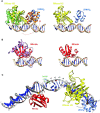
Model of UvrC binding to DNA. (A) Model of UvrC-C (yellow and blue) binding to DNA (right) assembled by overlaying (i) the RNase H2 domain of UvrC-C (yellow) onto Bacillus halodurans RNase H domain (green) bound to an RNA/DNA hybrid (PDB: 1ZBI) and, (ii) the (HhH)2 domain (blue) of UvrC-C onto E. coli RuvA (orange) bound to dsDNA (PDB: 1C7Y) as shown in the left panel. To achieve this DNA-bound conformation, the (HhH)2 motif has to undergo a near 180° anti-clockwise rotation (red dashed arrow) relative to the long α4 helix of the RNase H domain. For clarity, the DNA and RNA/DNA duplexes from the homologous structures are not shown. (B) Model of NEndo (red) binding to DNA (right) assembled by overlaying UvrC-NEndo onto E. coli GIY-YIG endonuclease R.Eco29kl (purple) bound to DNA (PDB: 3NIC) as shown in the left panel. (C) Model of NEndo (red) and UvrC-C (yellow and blue) binding to partially unwound UvrB-bound (not shown for clarity) pre-incision DNA extracted from the co-crystal structure of the B. caldotenax UvrB-DNA pre-incision complex (PDB: 6O8F) (73), which we extended on either end with standard B-form duplexed DNA. The flipped out damaged nucleotide is circled in green and the two endonuclease domains are positioned so as to perform their respective incision reactions (indicated with orange arrows) 4 nt downstream (NEndo) and 7 nt upstream (RNase H2) of the damaged site.
Figure 7.
Figure 7.
Proposed models of UvrC activation upon UvrB and DNA binding and formation of the UvrB/UvrC pre-incision complex. Alone, UvrC adopts a closed, inactive state (left panel) where access to the active sites of its two endonuclease domains is blocked. Upon binding to UvrB and DNA, UvrC is proposed to undergo a major conformational change, permitted by the flexible linkers between its domains, to adopt a more open and extended conformation in which it can both interact with the C-terminal domain of UvrB (UvrB-CTD) and with the DNA (middle and right panels). Full activation of UvrC additionally requires the partial unwinding of the DNA duplex in the vicinity of the damaged nucleotide by the dual action of UvrA and UvrB. In our present model (right) of the UvrB/UvrC pre-incision complex, although the exact positioning of the central domains of UvrC, and notably the RNase H1 domain, are still only speculative, both the N- and C-terminal regions of UvrC may be in contact with the UvrB-DNA pre-incision complex.

References

    1. Kisker C., Kuper J., Van Houten B.. Prokaryotic nucleotide excision repair. Cold Spring Harb. Perspect. Biol. 2013; 5:a012591. - PMC - PubMed
    1. Kad N.M., Van Houten B.. Dynamics of lesion processing by bacterial nucleotide excision repair proteins. Prog. Mol. Biol. Transl. Sci. 2012; 110:1–24. - PMC - PubMed
    1. Truglio J.J., Croteau D.L., Van Houten B., Kisker C.. Prokaryotic nucleotide excision repair: the UvrABC system. Chem. Rev. 2006; 106:233–252. - PubMed
    1. Jaciuk M., Swuec P., Gaur V., Kasprzak J.M., Renault L., Dobrychłop M., Nirwal S., Bujnicki J.M., Costa A., Nowotny M.. A combined structural and biochemical approach reveals translocation and stalling of UvrB on the DNA lesion as a mechanism of damage verification in bacterial nucleotide excision repair. DNA Repair (Amst.). 2020; 85:102746. - PMC - PubMed
    1. Pakotiprapha D., Jeruzalmi D.. Small-angle X-ray scattering reveals architecture and A(2)B(2) stoichiometry of the UvrA-UvrB DNA damage sensor. Proteins. 2013; 81:132–139. - PubMed

Publication types