. 2023 May:242:112167.
doi: 10.1016/j.jinorgbio.2023.112167. Epub 2023 Feb 26.

Revealing substrate-induced structural changes in active site of human CYP51 in the presence of its physiological substrates

Affiliations

Revealing substrate-induced structural changes in active site of human CYP51 in the presence of its physiological substrates

Yuanqi Jing et al. J Inorg Biochem. 2023 May.

Abstract

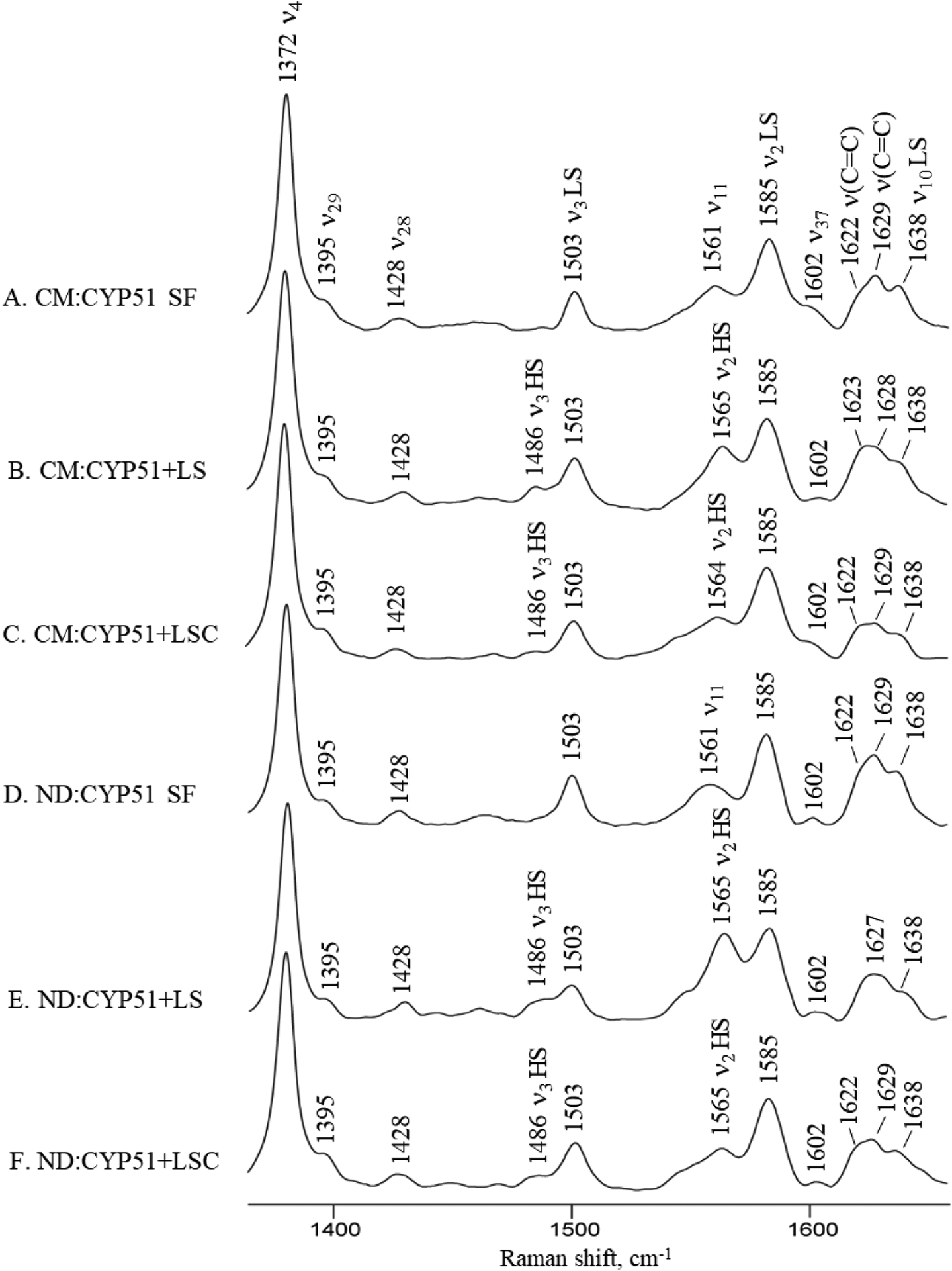
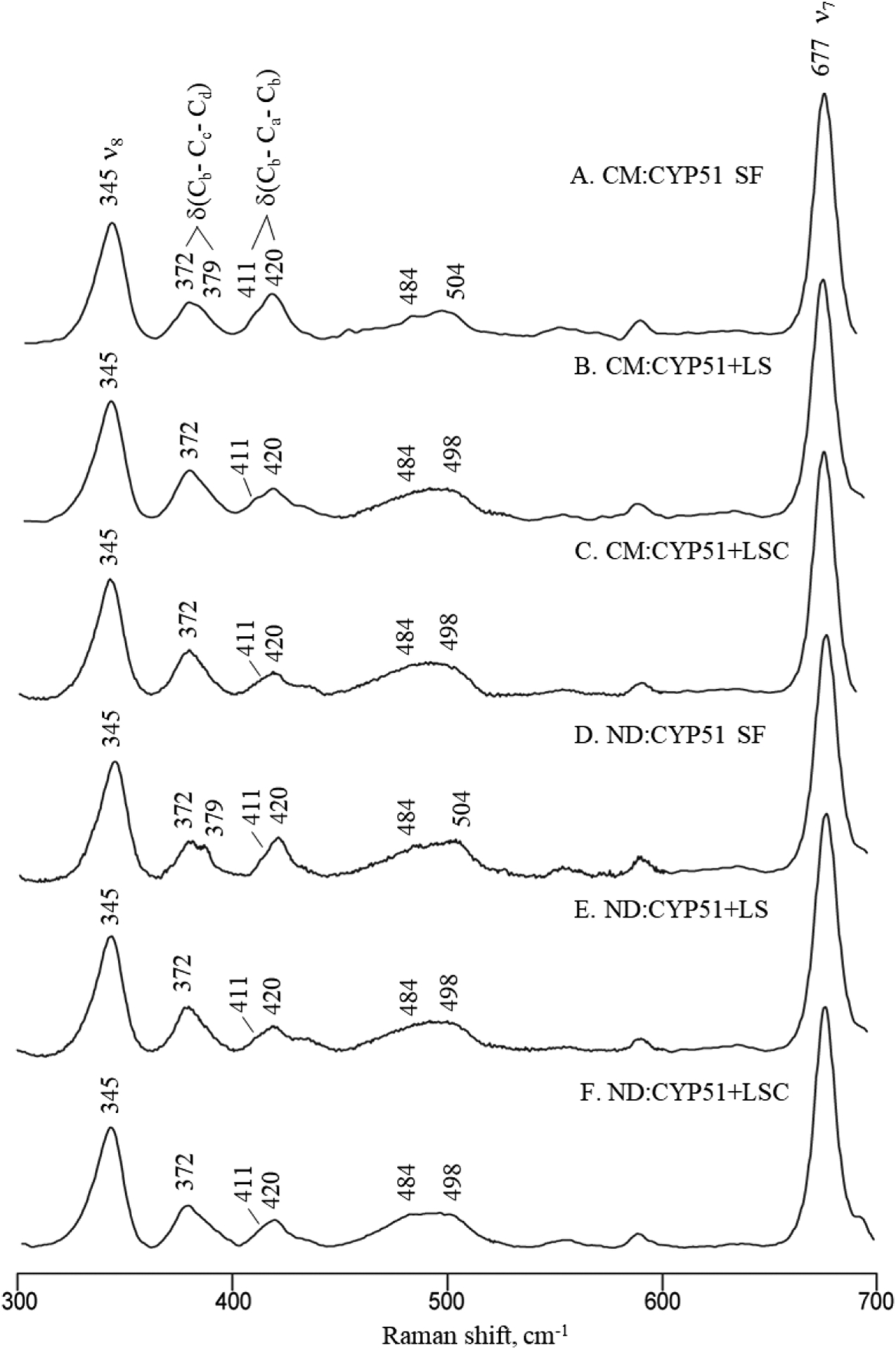
The human sterol 14α-demethylases (CYP51, CYP is an abbreviation for cytochrome P450) catalyze three-step oxidative removal of 14α-methyl group of lanosterol by first forming an alcohol, then an aldehyde, and finally conducting a CC bond cleavage reaction. This present study utilizes a combination of Resonance Raman spectroscopy and Nanodisc technology to probe the active site structure of CYP51 in the presence of its hydroxylase and lyase substrates. Ligand-binding induced partial low-to-high-spin conversion is observed by applying electronic absorption spectroscopy and Resonance Raman (RR) spectroscopy. This low degree of spin conversion of CYP51 is contributed by the retention of the water ligand coordinated to the heme iron as well as direct interaction between the hydroxyl group of lyase substrate and the iron center. No significant changes in active site structure are found between detergent-stabilized CYP51 and nanodisc-incorporated CYP51, nevertheless, it is demonstrated that nanodisc-incorporated assemblies provide much more well-defined active site RR spectroscopic responses, which induces a larger conversion from low-to-high-spin state in presence of the substrates. Moreover, a positive polar environment around the exogenous diatomic ligand is detected, providing insight into the mechanism of this essential CC bond cleavage reaction.

Keywords: Human CYP51; Nanodisc technology; Resonance Raman spectroscopy.


Conflict of interest statement

Declaration of Competing Interest The authors declare that they have no known competing financial interests or personal relationships that could have appeared to influence the work reported in this paper.

Figures

Figure 1.
Figure 1.
The three-step oxidative removal of 14α-methyl group of lanosterol.
Figure 2.
Figure 2.
Catalytic cycle of P450s. The active species that are proposed to be involved in hydroxylase and lyase activity are in the red bracket.
Figure 3.
Figure 3.
High-frequency RR data of ferric human CYP51. The top three traces are spectra of detergent-solubilized CYP51 samples: substrate-free (A), LS-bound (B), and LSC-bound (C). The bottom three traces are spectra of substrate-free (D), LS-bound (E), and LSC-bound (F) nanodisc-incorporated CYP51 samples. The bands in ν3 regions were resolved using peak fitting program, which was shown in Supporting information S6.
Figure 4.
Figure 4.
Low-frequency RR data of ferric human CYP51. The top three traces are spectra of detergent-solubilized CYP51 samples: substrate-free (A), LS-bound (B), and LSC-bound (C). The bottom three traces are spectra of substrate-free (D), LS-bound (E), and LSC-bound (F) nanodisc samples. The broad bands were resolved using peak fitting program, which was shown in Supporting information S7.
Figure 5.
Figure 5.
Low-frequency (left panel) and high-frequency (right panel) RR spectra of ferrous CO adducts of substrate-free ND: CYP51 (A), LS-bound ND: CYP51 (B), and LSC-bound ND: CYP51.
Figure 6.
Figure 6.
Inverse correlation plot showing ν(Fe-C)/ν(C-O) points of CYP51 and other P450s. The numbers correspond to the proteins and corresponding references are listed in supporting information table S1. The green squares show points for CYP101, the blue triangles represent points for CYP17A1, and the red circles show points for CYP51.
Figure 7.
Figure 7.
Proposed active site structure of CYP51A1 with presence of LS (a) and LSC (b).
Figure 8.
Figure 8.
Proposed active site structure of ferrous CO complex of CYP51. (a) substrate-free; (b) lanosterol bound; (c) aldehyde bound; (d) gem-diol bound.

References

    1. Lepesheva GI; Podust LM; Bellamine A; Waterman MR Folding Requirements Are Different between Sterol 14α-Demethylase (CYP51) from Mycobacterium Tuberculosis and Human or Fungal Orthologs. J. Biol. Chem 2001, 276, 28413–28420. 10.1074/jbc.M102767200. - DOI - PubMed
    1. Lepesheva GI; Waterman MR CYP51 - the Omnipotent P450. Mol. Cell. Endocrinol 2004, 215, 165–170. 10.1016/j.mce.2003.11.016. - DOI - PubMed
    1. Lepesheva GI; Virus C; Waterman MR Conservation in the CYP51 Family. Role of the B′ Helix/BC Loop and Helices F and G in Enzymatic Function. Biochemistry 2003, 42, 9091–9101. 10.1021/bi034663f. - DOI - PubMed
    1. Hargrove TY; Friggeri L; Wawrzak Z; Sivakumaran S; Yazlovitskaya EM; Hiebert SW; Guengerich FP; Waterman MR; Lepesheva GI Human Sterol 14α-Demethylase as a Target for Anticancer Chemotherapy: Towards Structure-Aided Drug Design. J. Lipid Res 2016, 57, 1552–1563. 10.1194/jlr.M069229. - DOI - PMC - PubMed
    1. Warrilow AG; Parker JE; Kelly DE; Kelly SL Azole Affinity of Sterol 14-Demethylase (CYP51) Enzymes from Candida Albicans and Homo Sapiens. Antimicrob. Agents Chemother 2013, 57, 1352–1360. 10.1128/AAC.02067-12. - DOI - PMC - PubMed

Publication types

LinkOut - more resources