. 2023 Apr:90:104499.
doi: 10.1016/j.ebiom.2023.104499. Epub 2023 Mar 2.

Neutrophil extracellular traps as a unique target in the treatment of chemotherapy-induced peripheral neuropathy

Affiliations

Neutrophil extracellular traps as a unique target in the treatment of chemotherapy-induced peripheral neuropathy

Chao-Yu Wang et al. EBioMedicine. 2023 Apr.

Abstract

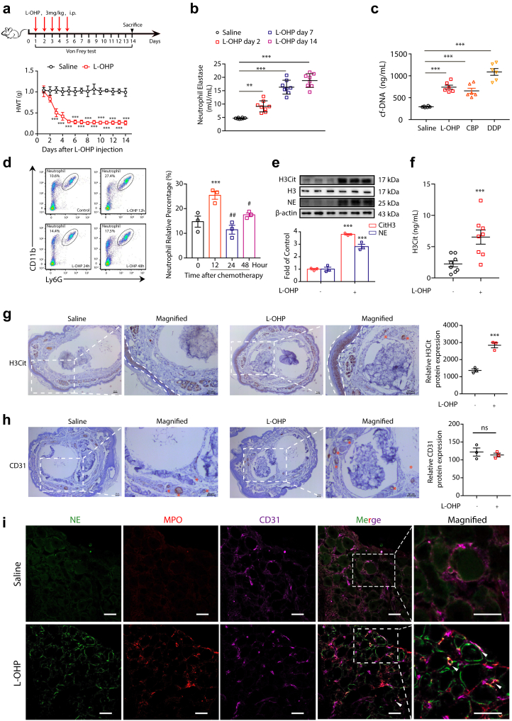
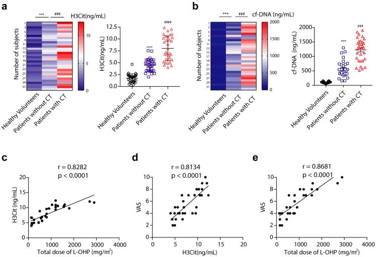
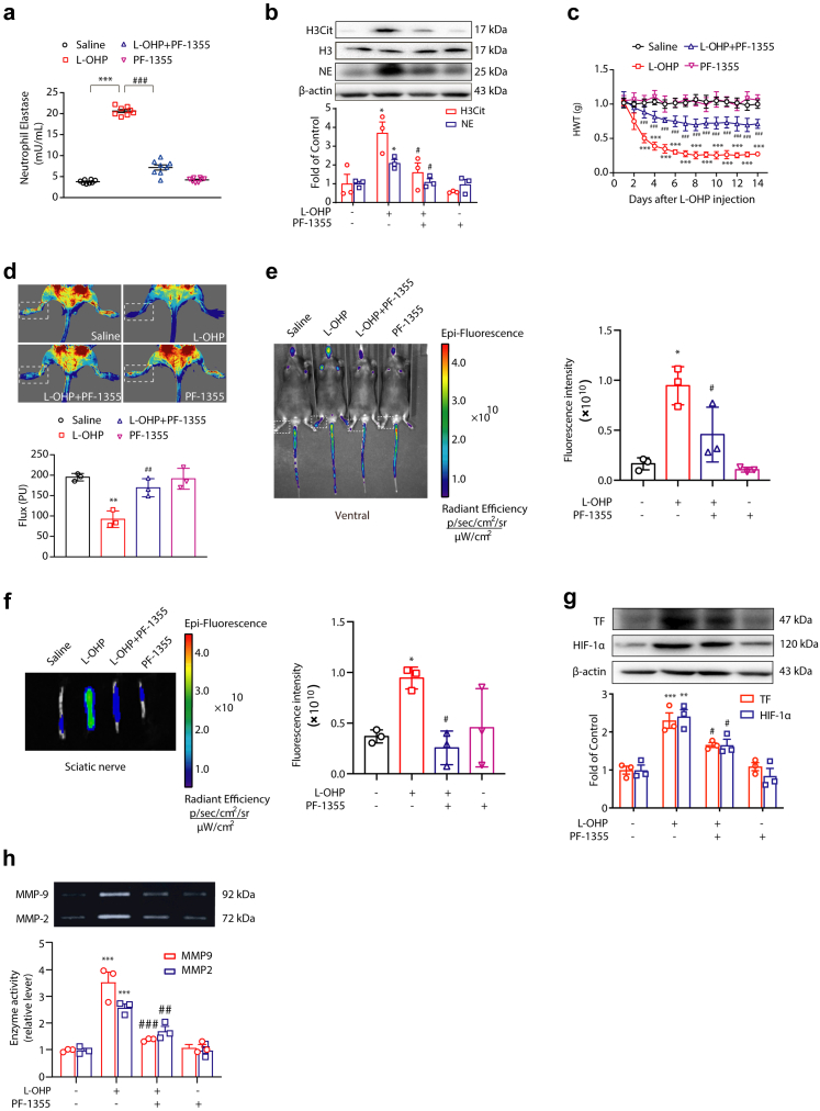
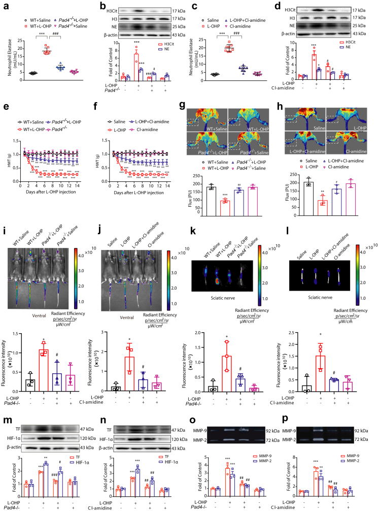
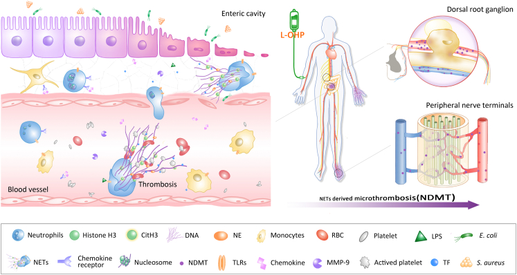
Background: Chemotherapy-induced peripheral neuropathy (CIPN) is a severe dose-limiting side effect of chemotherapy and remains a huge clinical challenge. Here, we explore the role of microcirculation hypoxia induced by neutrophil extracellular traps (NETs) in the development of CIPN and look for potential treatment.

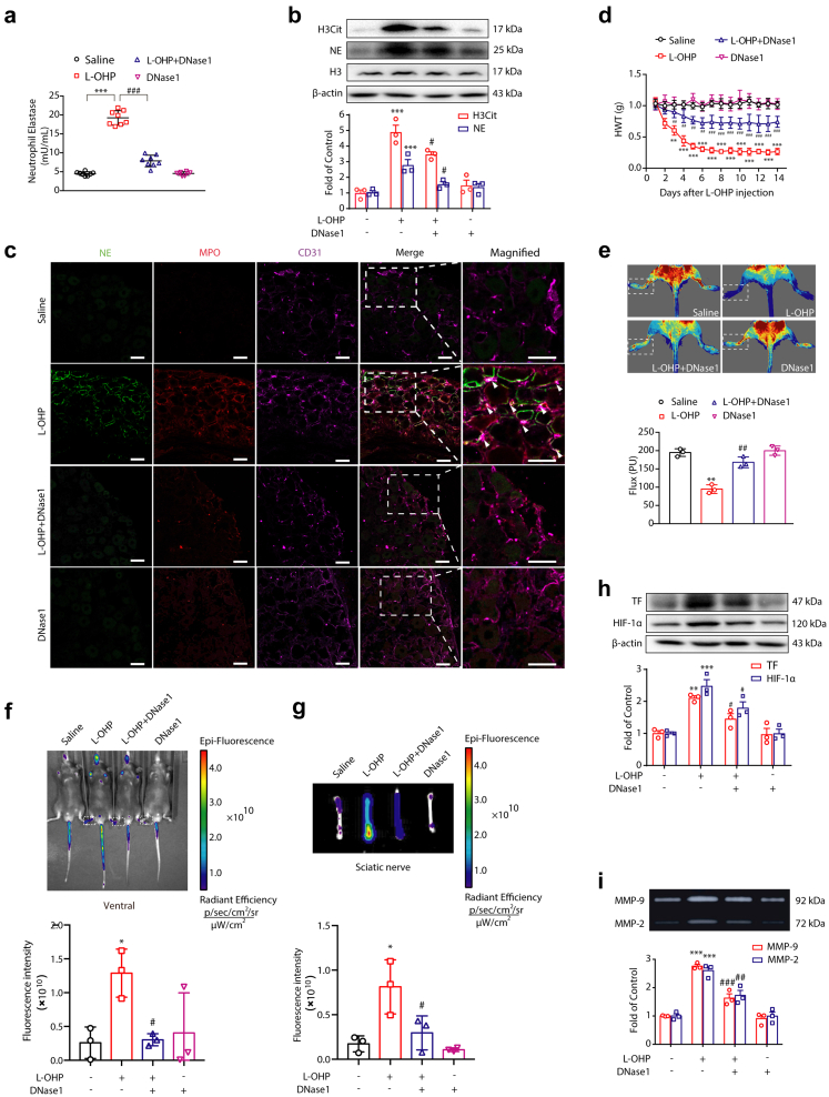
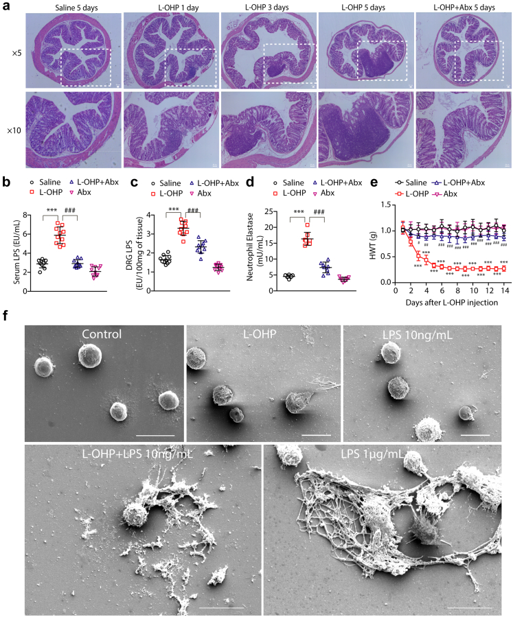
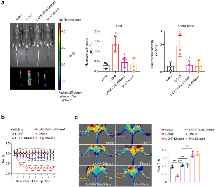
Methods: The expression of NETs in plasma and dorsal root ganglion (DRG) are examined by ELISA, IHC, IF and Western blotting. IVIS Spectrum imaging and Laser Doppler Flow Metry are applied to explore the microcirculation hypoxia induced by NETs in the development of CIPN. Stroke Homing peptide (SHp)-guided deoxyribonuclease 1 (DNase1) is used to degrade NETs.

Findings: The level of NETs in patients received chemotherapy increases significantly. And NETs accumulate in the DRG and limbs in CIPN mice. It leads to disturbed microcirculation and ischemic status in limbs and sciatic nerves treated with oxaliplatin (L-OHP). Furthermore, targeting NETs with DNase1 significantly reduces the chemotherapy-induced mechanical hyperalgesia. The pharmacological or genetic inhibition on myeloperoxidase (MPO) or peptidyl arginine deiminase-4 (PAD4) dramatically improves microcirculation disturbance caused by L-OHP and prevents the development of CIPN in mice.

Interpretation: In addition to uncovering the role of NETs as a key element in the development of CIPN, our finding provides a potential therapeutic strategy that targeted degradation of NETs by SHp-guided DNase1 could be an effective treatment for CIPN.

Funding: This study was funded by the National Natural Science Foundation of China81870870, 81971047, 81773798, 82271252; Natural Science Foundation of Jiangsu ProvinceBK20191253; Major Project of "Science and Technology Innovation Fund" of Nanjing Medical University2017NJMUCX004; Key R&D Program (Social Development) Project of Jiangsu ProvinceBE2019732; Nanjing Special Fund for Health Science and Technology DevelopmentYKK19170.

Keywords: Chemotherapy-induced peripheral neuropathy; Microcirculatory disturbance; Neutrophil extracellular traps; SHp-DNase1.


Conflict of interest statement

Declaration of interests The authors declare that they have no competing interests.

Figures

Fig. 1
Fig. 1
Chemotherapy triggers NETs formation. (a) Mice were intraperitoneally injected with L-OHP (3 mg/kg) for five consecutive days to induce mechanical hyperalgesia. The mechanical pain threshold was tested for 14 days by Von Frey Hairs. Significant differences were revealed following one-way ANOVA (n = 8, ∗∗∗P < 0.001 vs. saline). Hindpaw mechanical withdrawal threshold (HWT) was examined at indicated time points after oxaliplatin therapy. (b) After intraperitoneal injection of L-OHP (3 mg/kg), the content of NETs in blood after 2, 7, and 14 days was evaluated by a NETosis Assay Kit. (c) After intraperitoneal injection of L-OHP (3 mg/kg) for 5 days, CBP (10 mg/kg) for 1, 3, and 7 days, and DDP (2.3 mg/kg) for 5 days, the content of cf-NDA in blood was evaluated by a Quant-iTPico green dsDNA kit. CBP= Carboplatin, DDP = Cisplatin. B and C Data were analysed by one-sample t test (∗∗∗P < 0.001 vs. saline). (d) 12, 24, and 48 h after a single intraperitoneal injection of L-OHP (3 mg/kg), the proportion of neutrophils in the peripheral blood of mice was evaluated by flow cytometry, and the neutrophils were labeled with triple positivity of CD45, CD11b, and Ly-6G. Data were analysed by one-way ANOVA (n = 3, ∗∗∗P < 0.001 vs. 0 h; #P < 0.05, ##P < 0.01 vs. 12 h). (e) The protein levels of H3Cit and NE in the DRG were evaluated on the 14th day by Western blot. Data were analysed by one-way ANOVA (n = 3, ∗∗∗P < 0.001 vs. saline). (f) The levels of H3Cit in the plasma were evaluated on the 14th day by H3Cit ELISA kit. Data were analyzed by one-sample t test (∗∗∗P < 0.001 vs. saline). (g) Representative microscopy images of hind paw cross-sections from mice treated with saline or L-OHP and stained with the NETs marker H3Cit. The zones of the dermis and subcutaneous tissue are labeled with dotted lines, and the arteries are labeled with asterisks. Scale bars, 50 μm. (h) Representative microscopy images of hind paw cross-sections from mice treated with saline or L-OHP and stained with CD31. Vessels were labeled with asterisks. Scale bars, 50 μm. (i) Representative confocal immunofluorescence microscopy images of DRG from mice treated with saline or L-OHP for 14 days and stained with NE (green), MPO (red) and CD31 (purple); arrowheads point to NETs in vessels. Scale bars, 50 μm.
Fig. 2
Fig. 2
NETs presents in the plasma of patients after chemotherapy. (a and b) The level of H3Cit and cf-DNA measured by H3Cit ELISA kit and Quant-iTPico green dsDNA assay in plasma from healthy volunteers and cancer patients who had or had not received chemotherapy (n = 10) Data was analysed by one-way ANOVA (n = 32 volunteers in each group; ∗∗∗P < 0.001 vs. healthy volunteers; ###P < 0.001 vs. patients without chemotherapy). (c) Correlation between H3Cit and the total dose of L-OHP were analyzed by linear regression analysis (n = 32 cancer patients receiving chemotherapy, r = 0.8282, P < 0.0001, linear regression analysis). (d) Correlation between VAS and H3Cit and the correlation in cancer patients after chemotherapy were analysed by linear regression analysis (n = 32 cancer patients with chemotherapy, r = 0.8134, P = 0.0020, linear regression analysis). (e) Correlation between VAS and total dose of L-OHP (n = 32 cancer patients receiving chemotherapy, r = 0.8681, P < 0.0001, linear regression analysis). cf-DNA = circulating free DNA; VAS = visual analogue scale.
Fig. 3
Fig. 3
DNase1 alleviates CIPN by degrading NETs. Mice were intraperitoneally injected with L-OHP (3 mg/kg) for five consecutive days to induce mechanical hyperalgesia. DNase1 (150 U, i.v.) was administered from the first day of modeling to the 14th day. (a) The level of NETs in plasma was measured on the 14th day by a NETosis Assay Kit (n = 8). (b) The protein levels of H3Cit and NE in the DRG were evaluated on the 14th day by Western blot (n = 3). (c) Representative confocal immunofluorescence microscopy images of DRG from mice stained with NE (green), MPO (red) and CD31 (purple); arrowheads point to NETs in vessels (n = 3). Scale bars, 50 μm. (d) The mechanical pain threshold was tested for 14 days by Von Frey Hairs (n = 8). (e) Blood flow in the lower limbs of mice was evaluated by a moorFLPI2 flow speckle hemodynamic meter on the 14th day (n = 3). (f and g) SHp conjugated with the fluorescent dye Cy5-NHS was injected into mice via the tail vein. The ischemic condition of the limbs (f) and sciatic nerves (g) was evaluated by the IVIS Spectrum small animal in vivo visible three-dimensional imaging system after 2 h of SHp-Cy5 injection on the 14th day (n = 3). (h) The protein levels of TF and HIF-1α (n = 3) in the DRG were evaluated by Western blot. (i) The activities of MMP-9 (n = 3) in the DRG were evaluated by gelatin zymography. a, b, e, h and i data were analysed by one-way ANOVA. d data was analysed by two-way ANOVA. ∗∗P < 0.01, ∗∗∗P < 0.001 vs. saline; #P < 0.05, ##P < 0.01, ###P < 0.001 vs. the L-OHP-treated group.
Fig. 4
Fig. 4
Chemotherapy induced gut microbe-derived NETs formation. Mice were intraperitoneally injected with L-OHP (3 mg/kg) for 5 days, and antibiotics were added into the water three weeks in advance, which was freely ingested by the mice. (a) The degree of intestinal barrier and inflammatory infiltration was evaluated by H&E staining (n = 3). Scale bar, 50 μm. (b and c) LPS levels in the serum of mice were evaluated by a Pierce Chromogenic Endotoxin Quant Kit (n = 10). (d) The content of NETs in blood plasma was evaluated by a NETosis Assay Kit (n = 8). (e) The mechanical pain threshold was tested for 14 days by Von Frey Hairs (n = 8). (f) NETs formation was examined by scanning electron microscopy following stimulation of neutrophils with LPS (10 ng/mL), L-OHP (10 μM), LPS (10 ng/mL) + L-OHP (10 μΜ) for 4 h. Cells treated with LPS (1 μg/mL) were used as a positive control (n = 3). Scale bar, 10 μm. b–d data were analysed by one-way ANOVA. e data was analysed by two-way ANOVA. ∗∗∗P < 0.001 vs. saline; #P < 0.05, ##P < 0.01, ###P < 0.001 vs. the L-OHP-treated group.
Fig. 5
Fig. 5
MPO is responsible for L-OHP-induced NETs formation. Mice were intraperitoneally injected with L-OHP (3 mg/kg) for 5 days. MPO inhibitor PF-1355 (50 mg/kg, i.p.) was administered one day before L-OHP administration until the end of 14 days. (a) The levels of NETs in plasma were evaluated on the 14th day by a NETosis Assay Kit (n = 8). (b) The protein levels of H3Cit and NE in the DRG were evaluated on the 14th day by Western blot (n = 3). (c) The mechanical pain threshold was tested for 14 days by Von Frey Hairs (n = 8). (d) Blood flow in the lower limbs of mice was evaluated by a moorFLPI2 flow speckle hemodynamic meter on the 14th day (n = 3). (e and f) SHp conjugated with the fluorescent dye Cy5-NHS was injected into mice via the tail vein. The ischemic condition of the limbs (e) and sciatic nerves (f) was evaluated by the IVIS Spectrum small animal in vivo visible three-dimensional imaging system after 2 h of SHp-Cy5 injection on the 14th day (n = 3). (g) The protein levels of TF and HIF-1α (n = 3) in the DRG were evaluated by Western blot. (h) The activities of MMP-9 (n = 3) in the DRG were evaluated by gelatin zymography. a, b, g and h data were analysed by one-way ANOVA. c data was analysed by two-way ANOVA. ∗P < 0.05, ∗∗P < 0.01, ∗∗∗P < 0.001 vs. saline; #P < 0.05, ##P < 0.01, ###P < 0.001 vs. the L-OHP-treated group.
Fig. 6
Fig. 6
PAD4 is responsible for L-OHP-induced NETs formation. Mice were intraperitoneally injected with L-OHP (3 mg/kg) for 5 days. Pad4−/− mice were used to investigate NETs formation induced by L-OHP. Cl-amidine (10 mg/kg, i.p.) was administered one day before L-OHP administration until the end of 14 days. (a and c) The levels of NETs in plasma were evaluated on the 14th day by a NETosis Assay Kit (n = 8). (b and d) The protein levels of H3Cit and NE in the DRG were evaluated on the 14th day by Western blot (n = 3). (e and f) The mechanical pain threshold was tested for 14 days by Von Frey Hairs (n = 8). (g and h) Blood flow in the lower limbs of mice was evaluated by a moorFLPI2 flow speckle hemodynamic meter on the 14th day (n = 3). (il) SHp conjugated with the fluorescent dye Cy5-NHS was injected into mice via the tail vein. The ischemic conditions of the limbs (i and j) and sciatic nerves (k and l) were evaluated by the IVIS Spectrum small animal in vivo visible three-dimensional imaging system after 2 h of SHp-Cy5 injection on the 14th day (n = 3). (m and n) The protein levels of TF and HIF-1α (n = 3) in the DRG were evaluated by Western blot. (o and p) The activities of MMP-9 (n = 3) in the DRG were evaluated by gelatin zymography. a–d, g, h, m–p data were analysed by one-way ANOVA. e and f data were analysed by two-way ANOVA. ∗∗P < 0.01, ∗∗∗P < 0.001 vs. saline; #P < 0.05, ##P < 0.01, ###P < 0.001 vs. L-OHP-treated group in a, b, e, g, m and o data. ∗P < 0.05, ∗∗P < 0.01, ∗∗∗P < 0.001 vs. saline; #P < 0.05, ##P < 0.01, ###P < 0.001 vs. WT + L-OHP group in c, d, f, h, n and p data.
Fig. 7
Fig. 7
SHp-DNase1 treatment prevents chemotherapy-induced mechanical hyperalgesia. Mice were intraperitoneally injected with L-OHP (3 mg/kg) for five consecutive days to induce mechanical hyperalgesia. (a) SHp-Cy5 was injected into mice via the tail vein. The ischemic condition of the limbs and sciatic nerves was evaluated by the IVIS Spectrum small animal in vivo visible three-dimensional imaging system after 2 h of SHp-Cy5 injection on the 14th day (n = 3). (b) DNase1 (150U, i.v.) or SHp-DNase1 (150U, i.v.) was administered from the first day of modeling to the 14th day. The mechanical pain threshold was tested for 14 days by Von Frey Hairs (n = 8). Data were analysed by two-way ANOVA. ∗∗P < 0.01, ∗∗∗P < 0.001 vs. saline; ##P < 0.01, ###P < 0.001 vs. the L-OHP-treated group. (c) DNase1 (150U, i.v.) or SHp-DNase1 (150U, i.v.) was administered from the first day of modeling to the 14th day. Blood flow in the lower limbs of mice was evaluated by a moor FLPI2 flow speckle hemodynamic meter on the 14th day (n = 3). Data were analysed by one-way ANOVA. ∗∗∗P < 0.001 vs. saline; #P < 0.05, ###P < 0.001 vs. the L-OHP-treated group. $P < 0.05 vs. L-OHP-DNase1-treated group.
Fig. 8
Fig. 8
Schematic illustration indicating that chemotherapy-induced NETs contribute to the development of CIPN by disturbing microcirculation. L-OHP leads to intestinal barrier destruction and inflammatory infiltration, and gut microbe-derived LPS triggers neutrophils to form NETs, which are subsequently released into the blood. NETs-mediated microcirculation disturbance causes ischemia and hypoxia in the DRG and limbs, which contributes to the progression of CIPN.

References

    1. Park S.B., Goldstein D., Krishnan A.V., et al. Chemotherapy-induced peripheral neurotoxicity: a critical analysis. CA Cancer J Clin. 2013;63:419–437. - PubMed
    1. Seretny M., Currie G.L., Sena E.S., et al. Incidence, prevalence, and predictors of chemotherapy-induced peripheral neuropathy: a systematic review and meta-analysis. Pain. 2014;155:2461–2470. - PubMed
    1. Wolf S., Barton D., Kottschade L., Grothey A., Loprinzi C. Chemotherapy-induced peripheral neuropathy: prevention and treatment strategies. Eur J Cancer. 2008;44:1507–1515. - PubMed
    1. LoMonaco M., Milone M., Batocchi A.P., Padua L., Restuccia D., Tonali P. Cisplatin neuropathy: clinical course and neurophysiological findings. J Neurol. 1992;239:199–204. - PubMed
    1. Lechner D., Weltermann A. Chemotherapy-induced thrombosis: a role for microparticles and tissue factor? Semin Thromb Hemost. 2008;34:199–203. - PubMed

MeSH terms