Review
. 2023 Apr:161:110757.
doi: 10.1016/j.ejrad.2023.110757. Epub 2023 Feb 25.

Prostate MRI and image Quality: It is time to take stock

Affiliations
Review

Prostate MRI and image Quality: It is time to take stock

Yue Lin et al. Eur J Radiol. 2023 Apr.

Abstract

Multiparametric magnetic resonance imaging (mpMRI) plays a vital role in prostate cancer diagnosis and management. With the increase in use of mpMRI, obtaining the best possible quality images has become a priority. The Prostate Imaging Reporting and Data System (PI-RADS) was introduced to standardize and optimize patient preparation, scanning techniques, and interpretation. However, the quality of the MRI sequences depends not only on the hardware/software and scanning parameters, but also on patient-related factors. Common patient-related factors include bowel peristalsis, rectal distension, and patient motion. There is currently no consensus regarding the best approaches to address these issues and improve the quality of mpMRI. New evidence has been accrued since the release of PI-RADS, and this review aims to explore the key strategies which aim to improve prostate MRI quality, such as imaging techniques, patient preparation methods, the new Prostate Imaging Quality (PI-QUAL) criteria, and artificial intelligence on prostate MRI quality.

Keywords: Diagnostic Imaging; Magnetic Resonance Imaging; Prostatic Neoplasms.


Conflict of interest statement

Declaration of Competing Interest The authors declare that they have no known competing financial interests or personal relationships that could have appeared to influence the work reported in this paper.

Figures
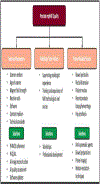
Figure 1.
Figure 1.
Factors that can impact prostate mpMRI quality.
Figure 2.
Figure 2.
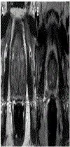
Axial T2W MRIs of a 63-year-old (serum PSA=9.5ng/ml) (A) and a 53-year-old (serum PSA=5ng/ml) (B) male obtained with endorectal coil. Use of endorectal coil resulted in motion related ghosting artifacts in both patients.
Figure 3.
Figure 3.
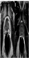
Axial T2W MRIs of a 60-year-old (serum PSA=4.8ng/ml) (A) and a 48-year-old (serum PSA=0.9ng/ml) (B) male obtained with endorectal coil. Use of endorectal coil resulted in deformations in the peripheral zone which is secondary to coil elements as a result of incorrect placement of the endorectal coils (arrows).
Figure 4.
Figure 4.
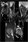
mpMRI of a 72-year-old male with a serum PSA of 6.9 ng/mL. Axial (A), sagittal (B), coronal T2WI (C), high b-value DWI (D), ADC map (E), and DCE MRI (F). Patient has a focal lesion in right mid peripheral zone which is hypointense on axial (A) and coronal (C) T2WIs, and has a focal contrast uptake on DCE (F). However, due to large amount of rectal gas, visible on axial and sagittal T2WIs (asterisk), high b-value and ADC map images are distorted and lesion cannot be visualized (arrowheads). Targeted biopsy of the lesion revealed a Gleason grade 7 (3+4) prostate adenocarcinoma.
Figure 5.
Figure 5.
mpMRI of a 55-year-old man with a serum PSA of 6.7 ng/mL. Axial (A), sagittal (B), coronal T2WI (C), high b-value DWI (D), and ADC map (E). Patient has a lesion in right apical-mid peripheral zone which is hypointense on axial (A) and coronal (C) T2WIs. Rectal gas, which can be seen on axial (A) and sagittal (B) T2WIs (asterisk), impacts the quality of high bvalue DWI (D) and ADC map (E) images which subsequently makes the intraprostatic lesion indistinguishable on these sequences (arrowheads). There is also an accompanying local distortion and shining in the posterolateral aspect of the left hemigland on high b-value DWI (D) (dashed arrow). Targeted biopsy of the lesion revealed a Gleason grade 7 (3+4) prostate adenocarcinoma.
Figure 6.
Figure 6.
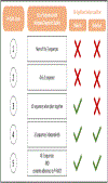
A diagram explaining how PI-QUAL scores are assigned and their implications.

References

    1. Kasivisvanathan V, Rannikko AS, Borghi M et al. (2018) MRI-Targeted or Standard Biopsy for Prostate-Cancer Diagnosis. N Engl J Med 378:1767–1777 - PMC - PubMed
    1. Ahdoot M, Wilbur AR, Reese SE et al. (2020) MRI-Targeted, Systematic, and Combined Biopsy for Prostate Cancer Diagnosis. N Engl J Med 382:917–928 - PMC - PubMed
    1. Ahmed HU, El-Shater Bosaily A, Brown LC et al. (2017) Diagnostic accuracy of multiparametric MRI and TRUS biopsy in prostate cancer (PROMIS): a paired validating confirmatory study. Lancet 389:815–822 - PubMed
    1. Greer MD, Shih JH, Lay N et al. (2019) Interreader Variability of Prostate Imaging Reporting and Data System Version 2 in Detecting and Assessing Prostate Cancer Lesions at Prostate MRI. AJR Am J Roentgenol. 10.2214/AJR.18.20536:1–8 - DOI - PMC - PubMed
    1. Turkbey B, Rosenkrantz AB, Haider MA et al. (2019) Prostate Imaging Reporting and Data System Version 2.1: 2019 Update of Prostate Imaging Reporting and Data System Version 2. Eur Urol 76:340–351 - PubMed