. 2023 Apr:117:109954.
doi: 10.1016/j.intimp.2023.109954. Epub 2023 Feb 27.

SARS-CoV-2 immune complex triggers human monocyte necroptosis

Affiliations

SARS-CoV-2 immune complex triggers human monocyte necroptosis

Leonardo Duarte Santos et al. Int Immunopharmacol. 2023 Apr.

Abstract

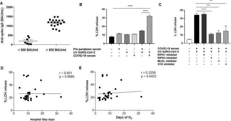
We analyzed the ability of severe acute respiratory syndrome coronavirus-2 (SARS-CoV-2) itself and SARS-CoV-2-IgG immune complexes to trigger human monocyte necroptosis. SARS-CoV-2 was able to induce monocyte necroptosis dependently of MLKL activation. Necroptosis-associated proteins (RIPK1, RIPK3 and MLKL) were involved in SARS-CoV-2N1 gene expression in monocytes. SARS-CoV-2 immune complexes promoted monocyte necroptosis in a RIPK3- and MLKL-dependent manner, and Syk tyrosine kinase was necessary for SARS-CoV-2 immune complex-induced monocyte necroptosis, indicating the involvement of Fcγ receptors on necroptosis. Finally, we provide evidence that elevated LDH levels as a marker of lytic cell death are associated with COVID-19 pathogenesis.

Keywords: COVD-19 pathogenesis; Fcγ receptors; MLKL; Monocyte; Necroptosis; SARS-CoV-2.


Conflict of interest statement

Declaration of Competing Interest The authors declare that they have no known competing financial interests or personal relationships that could have appeared to influence the work reported in this paper.

Figures

Fig. 1
Fig. 1
SARS-CoV-2 promotes human monocyte necroptosis. A) Human monocytes (3x10⁵/well) were treated with NEC-1s (30 μM), GW42X (2 μM), or NSA (2 μM) for 1 h prior to SARS-CoV-2 (MOI 0.5) infection for 24 h. Cell death was assessed by LDH release in cell supernatants and expressed as % LDH release. B) Human monocytes (3x10⁵/well) were treated with NEC-1s (30 μM), GW42X (2 μM), or NSA (2 μM) for 1 h prior to SARS-CoV-2 (MOI 0.5) infection for 24 h. RNA was harvested, and SARS-CoV-2 N1 mRNA expression was quantified by real-time PCR using the 2-ΔCt method. Data are representative of at least two independent experiments performed in triplicate and expressed as mean ± SEM. Data were analyzed with one-way ANOVA with Tukey’s post hoc test. *** p ≤ 0.001; **** p ≤ 0.0001.
Fig. 2
Fig. 2
SARS-CoV-2–IgG immune complex triggers RIPK3- and MLKL-mediated necroptosis in human monocytes dependently of Fcγ receptors. A) The concentration of IgG antibodies against the S1 antigen of SARS-CoV-2 was measured in the serum of COVID-19 patients using ELISA and expressed as BAU/mL. B) Human monocytes were stimulated with either UV-SARS-CoV-2, pre-pandemic serum, COVID-19 serum, the mixture of UV-SARS-CoV-2 + pre-pandemic serum, UV-SARS-CoV-2 immune complexes (UV-SARS-CoV-2 + COVID-19 serum), or media only for 24 h. Afterwards, cell death was assessed by LDH release in cell supernatants and expressed as % LDH release. C) Monocytes were treated with RIPK1 inhibitor (NEC-1s; 30 μM), RIPK3 inhibitor (GW42X; 2 μM), MLKL inhibitor (NSA; 2 μM), or Syk tyrosine kinase inhibitor (piceatannol; 20 μM) for 1 h prior to stimulation with UV-SARS-CoV-2 immune complexes (UV-SARS-CoV-2 + COVID-19 serum). Afterwards, cell death was assessed by LDH release in cell supernatants. D) Spearman linear correlation between LDH release levels and length of hospital stay of COVID-19 patients. E) Spearman linear correlation between LDH release levels and time of O2 need of COVID-19 patients. Data are representative of at least two independent experiments performed in triplicate and expressed as mean ± SEM. Data were analyzed with one-way ANOVA with Tukey’s post hoc test (B,C). ** p ≤ 0.01; *** p ≤ 0.001; **** p ≤ 0.0001.

References

    1. Knoll R., Schultze J.L., Schulte-Schrepping J. Monocytes and Macrophages in COVID-19. Front. Immunol. 2021;12 - PMC - PubMed
    1. Meidaninikjeh S., Sabouni N., Marzouni H.Z., Bengar S., Khalili A., Jafari R. Monocytes and macrophages in COVID-19: Friends and foes. Life Sci. 2021;269 - PMC - PubMed
    1. Frank D., Vince J.E. Pyroptosis versus necroptosis: similarities, differences, and crosstalk. Cell Death Differ. 2019;26(1):99–114. - PMC - PubMed
    1. Wu C., Chen X., Cai Y., Xia J., Zhou X., Xu S., Huang H., Zhang L., Zhou X., Du C., Zhang Y., Song J., Wang S., Chao Y., Yang Z., Xu J., Zhou X., Chen D., Xiong W., Xu L., Zhou F., Jiang J., Bai C., Zheng J., Song Y. Risk Factors Associated With Acute Respiratory Distress Syndrome and Death in Patients With Coronavirus Disease 2019 Pneumonia in Wuhan, China. JAMA Intern. Med. 2020;180(7):934–943. - PMC - PubMed
    1. Karki R., Sharma B.R., Tuladhar S., Williams E.P., Zalduondo L., Samir P., Zheng M., Sundaram B., Banoth B., Malireddi R.K.S., Schreiner P., Neale G., Vogel P., Webby R., Jonsson C.B., Kanneganti T.D. Synergism of TNF-alpha and IFN-gamma Triggers Inflammatory Cell Death, Tissue Damage, and Mortality in SARS-CoV-2 Infection and Cytokine Shock Syndromes. Cell. 2021;184(1):149–168 e17. - PMC - PubMed

MeSH terms

Substances