. 2023 Apr;103(4):100036.
doi: 10.1016/j.labinv.2022.100036. Epub 2023 Jan 10.

Intestinal Epithelial Digestive, Transport, and Barrier Protein Expression Is Increased in Environmental Enteric Dysfunction

Affiliations

Intestinal Epithelial Digestive, Transport, and Barrier Protein Expression Is Increased in Environmental Enteric Dysfunction

Shabnam Abtahi et al. Lab Invest. 2023 Apr.

Abstract

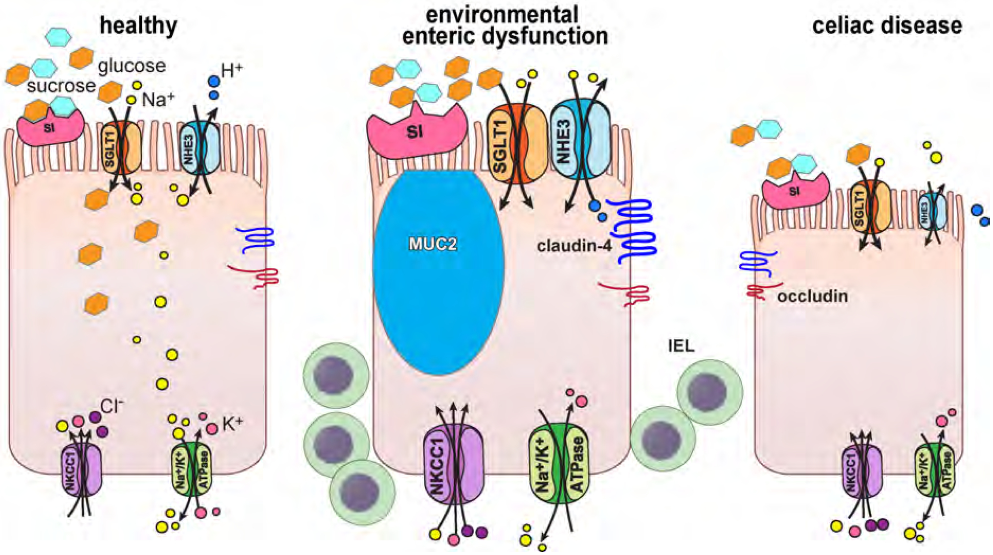
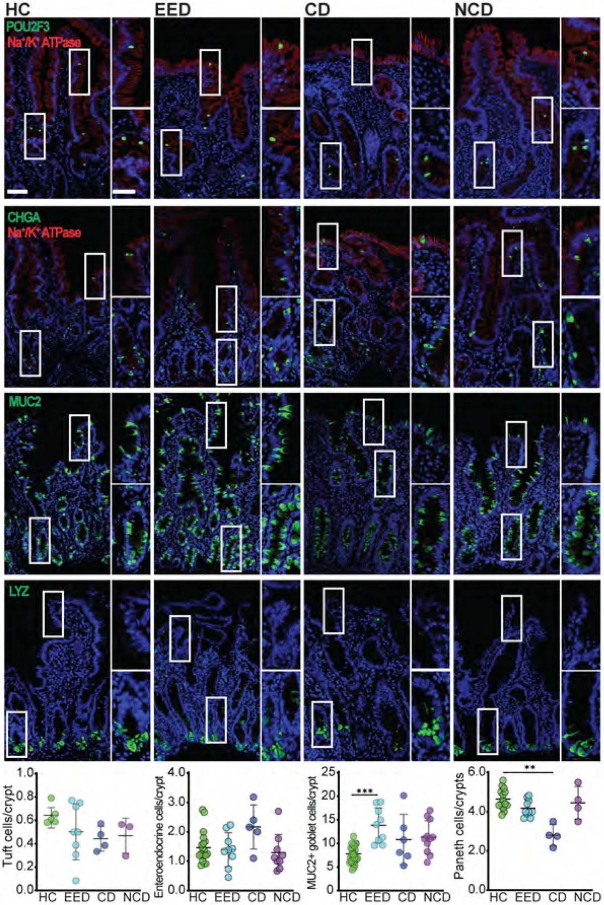
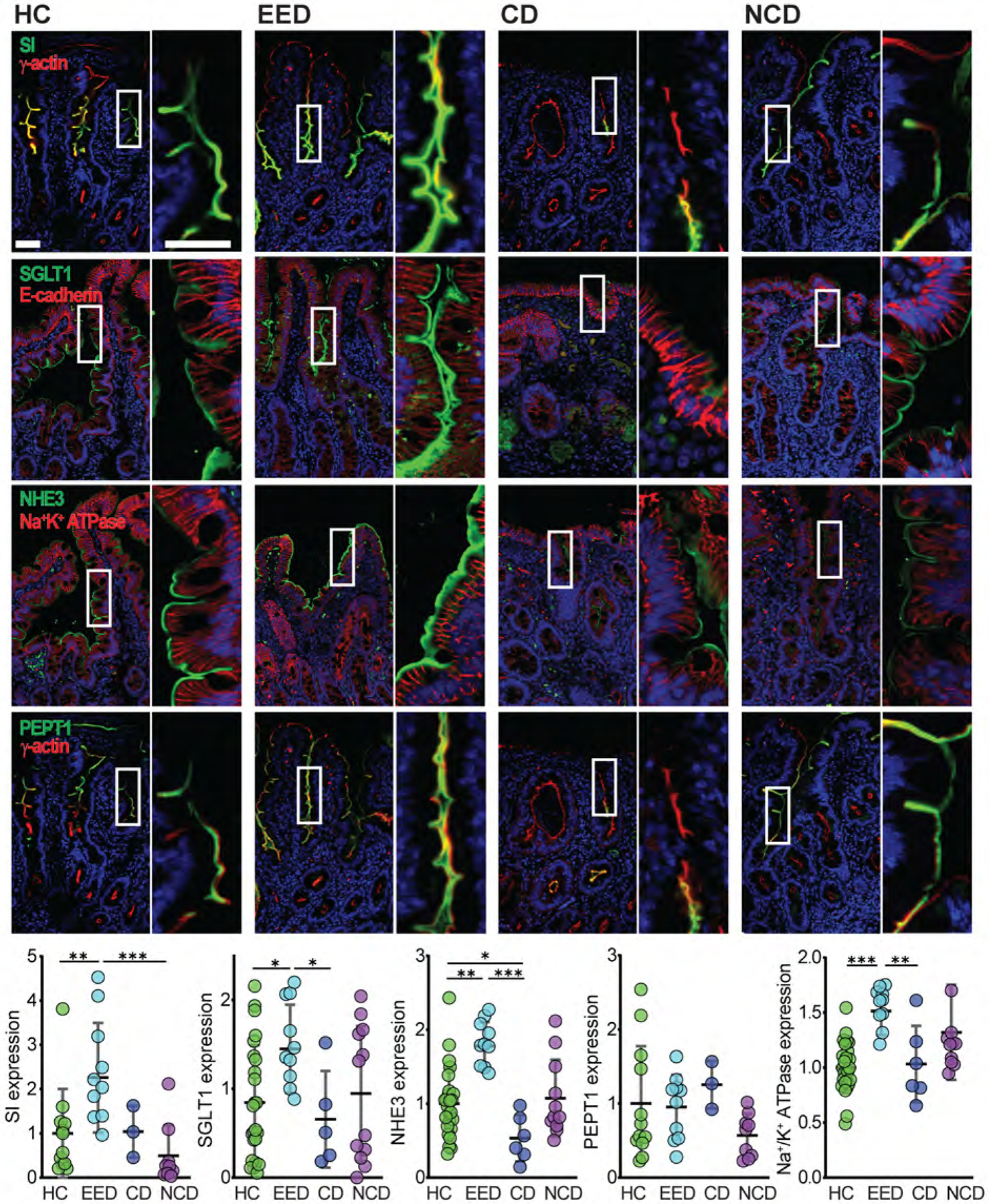
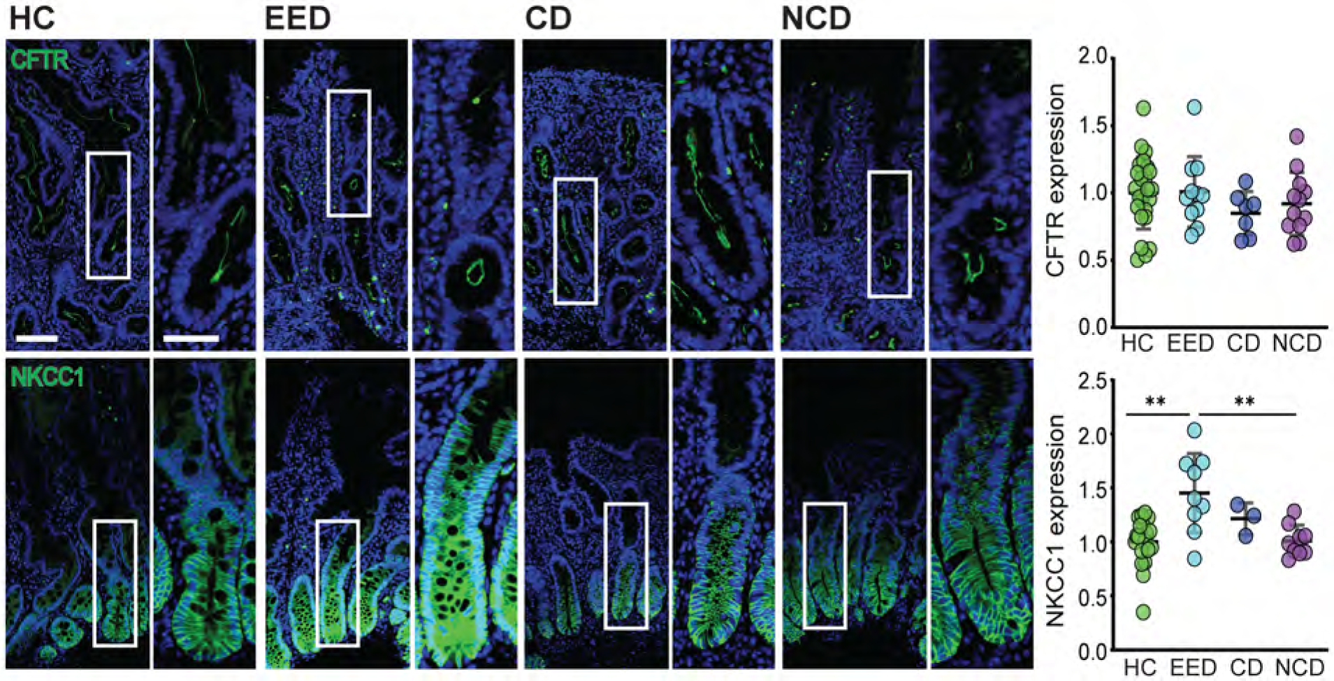
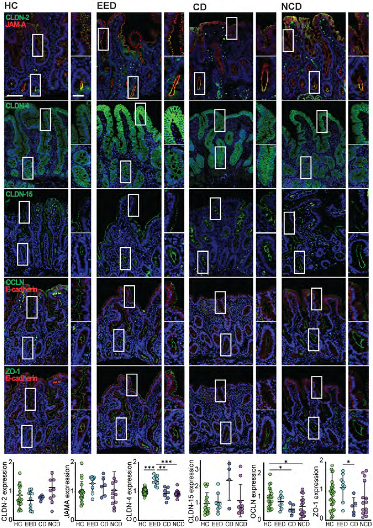
Environmental enteric dysfunction (EED) is characterized by malabsorption and diarrhea that result in irreversible deficits in physical and intellectual growth. We sought to define the expression of transport and tight junction proteins by quantitative analysis of duodenal biopsies from patients with EED. Biopsies from Pakistani children with confirmed EED diagnoses were compared to those from age-matched North American healthy controls, patients with celiac disease, and patients with nonceliac disease with villous atrophy or intraepithelial lymphocytosis. Expression of brush border digestive and transport proteins and paracellular (tight junction) proteins was assessed by quantitative multiplex immunofluorescence microscopy. EED was characterized by partial villous atrophy and marked intraepithelial lymphocytosis. Epithelial proliferation and enteroendocrine, tuft, and Paneth cell numbers were unchanged, but there was significant goblet cell expansion in EED biopsies. Expression of proteins involved in nutrient and water absorption and that of the basolateral Cl- transport protein NKCC1 were also increased in EED. Finally, the barrier-forming tight junction protein claudin-4 (CLDN4) was significantly upregulated in EED, particularly within villous enterocytes. In contrast, expression of CFTR, CLDN2, CLDN15, JAM-A, occludin, ZO-1, and E-cadherin was unchanged. Upregulation of a barrier-forming tight junction protein and brush border and basolateral membrane proteins that support nutrient and water transport in EED is paradoxical, as their increased expression would be expected to be correlated with increased intestinal barrier function and enhanced absorption, respectively. These data suggest that EED activates adaptive intestinal epithelial responses to enhance nutrient absorption but that these changes are insufficient to restore health.

Keywords: enteropathy; malabsorption; stunted growth; tight junction; tropical sprue.


Conflict of interest statement

Competing interests: JRT is a founder of Thelium Therapeutics and a consultant for Entrinsic and Kallyope. No other authors have competing interests.

Figures

Figure 1.
Figure 1.. EED is characterized by modest villus blunting and marked intraepithelial lymphocytosis.
Villus height was significantly reduced in children with EED or celiac disease (CD) relative to healthy controls (HC) and non-celiac disease (NCD) subjects. Villous atrophy in celiac disease was significantly more severe than in EED, but numbers of CD3+ intraepithelial lymphocytes (IELs) were more prominently increased in EED. Proliferation, assessed as numbers of Ki67-positive cells, was increased in celiac disease but unchanged in EED. Numbers of cleaved caspase 3-positive cells were similar in all groups. Bars = 50 μm, 20 μm. *, P < 0.05; **, P < 0.01; ***, P < 0.001.
Figure 2.
Figure 2.. Goblet cell populations are expanded in EED.
Numbers of POU2F3-positive tuft cells and chromogranin A-positive (CHGA) enteroendocrine cells were similar in all groups. MUC2-positive goblet cell numbers were increased in EED relative to all other groups. Data shown are per crypt unit. When quantified per 100 epithelial cells, MUC2-positive goblet cells numbered 14.7±2.2; 19.8±3.4; 16.5±1.5; and 20.0±4.3 for HC, EED, CD, and NCD groups, respectively. MUC2-positive goblet cell numbers in villous atrophy and intraepithelial lymphocytosis groups were 17.0±3.8; and 23.1±2.0, respectively. Goblet cell numbers were significantly increased in EED, NCD, and villous atrophy groups relative to HC. Loss of lysozyme-positive Paneth cells was evident in celiac disease. Bars = 50 μm and 20 μm. *, P < 0.05; **, P < 0.01; ***, P < 0.001.
Figure 3.
Figure 3.. Brush border proteins involved in nutrient and water absorption are upregulated in EED.
Expression of the brush border proteins sucrase-isomaltase (SI), the Na+-glucose cotransporter SGLT1, and the Na+/H+ exchanger NHE3 (SLC9A3), were all increased in EED, as was Na+/K+ATPase. Conversely, NHE3 was reduced in celiac disease. PEPT1 expression was not different in any group. Bars = 50 μm and 20 μm. *, P < 0.05; **, P < 0.01; ***, P < 0.001.
Figure 4.
Figure 4.. Basolateral Cl transporter expression is upregulated in EED.
CFTR expression was unchanged in EED. In contrast, expression of the basolateral Cl transporter NKCC1 was increased. Bars = 50 μm and 20 μm. *, P < 0.05; **, P < 0.01; ***, P < 0.001.
Figure 5.
Figure 5.. Expression of barrier-forming claudin-4 increased in EED.
Among the tight junction proteins examined, only claudin-4 (CLDN4) displayed EED-associated expression changes. Increased claudin-4 expression was particularly evident in the upper half of villi. In contrast, expression of the pore-forming claudins 2 (CLDN2) and 15 (CLDN15) as well as the leak pathway regulatory proteins occludin (OCLN), ZO-1, and JAM-A were unchanged. Similarly, E-cadherin expression was unchanged in EED. Occludin expression was reduced in celiac disease. Bars = 50 μm and 20 μm. *, P < 0.05; **, P < 0.01; ***, P < 0.001.
Figure 6.
Figure 6.. Comparisons of protein expression in biopsies from healthy subjects and those with EED or celiac disease.
The cell on the left represents healthy physiology. Sucrase-isomaltase (SI) breaks sucrose into glucose and fructose. Glucose is transported across the apical brush border by the Na+-glucose cotransporter, SGLT1 (SLC5A1). Glucose is then used as an energy source by the cell or exits the cell via basolateral GLUT2 (SLC2A2, not shown). Similarly, NHE3 (SLC9A3) takes advantage of the extracellular to intracellular Na+ gradient to transport H+ across the apical membrane from the cytoplasm to the lumen. In both cases, Na+ is then pumped across the basolateral membrane by Na+/K+ATPase. NKCC1 (SLC12A2), also located on the basolateral membrane, transports Na+. K+, and Cl into the cell, thereby providing the driving force for apical Cl secretion. SI, SGLT1, NHE3, NKCC1, and Na+/K+ATPase are all upregulated in EED, as is the barrier-forming tight junction protein claudin-4. Increased numbers of MUC2-positive goblet cells and intraepithelial lymphocytes (IEL) are also present. Although, like EED, intraepithelial lymphocytes are increased in celiac disease, NHE3 and occludin are downregulated on a per-cell basis. The loss of absorptive epithelial cells in celiac disease due to near-total villous atrophy results in reduced overall absorptive protein expression.

References

    1. Lin A, Ali S, Arnold BF, et al. Effects of Water, Sanitation, Handwashing, and Nutritional Interventions on Environmental Enteric Dysfunction in Young Children: A Cluster-randomized, Controlled Trial in Rural Bangladesh. Clin Infect Dis 2020; 70:738–747. - PubMed
    1. Keusch GT, Denno DM, Black RE, et al. Environmental enteric dysfunction: pathogenesis, diagnosis, and clinical consequences. Clin Infect Dis 2014; 59 Suppl 4:S207–12. - PMC - PubMed
    1. Semba RD, Shardell M, Trehan I, et al. Metabolic alterations in children with environmental enteric dysfunction. Sci Rep 2016; 6:28009. - PMC - PubMed
    1. Haberman Y, Iqbal NT, Ghandikota S, et al. Mucosal Genomics Implicate Lymphocyte Activation and Lipid Metabolism in Refractory Environmental Enteric Dysfunction. Gastroenterology 2021; 160:2055–2071 e0. - PMC - PubMed
    1. Amadi B, Besa E, Zyambo K, et al. Impaired Barrier Function and Autoantibody Generation in Malnutrition Enteropathy in Zambia. EBioMedicine 2017; 22:191–199. - PMC - PubMed

Publication types

Substances