. 2023 May 1:200:47-58.
doi: 10.1016/j.freeradbiomed.2023.03.001. Epub 2023 Mar 2.

Activation of neuronal NADPH oxidase NOX2 promotes inflammatory neurodegeneration

Affiliations

Activation of neuronal NADPH oxidase NOX2 promotes inflammatory neurodegeneration

Dezhen Tu et al. Free Radic Biol Med. .

Abstract

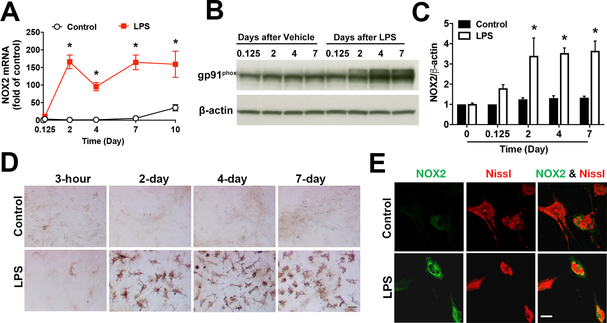
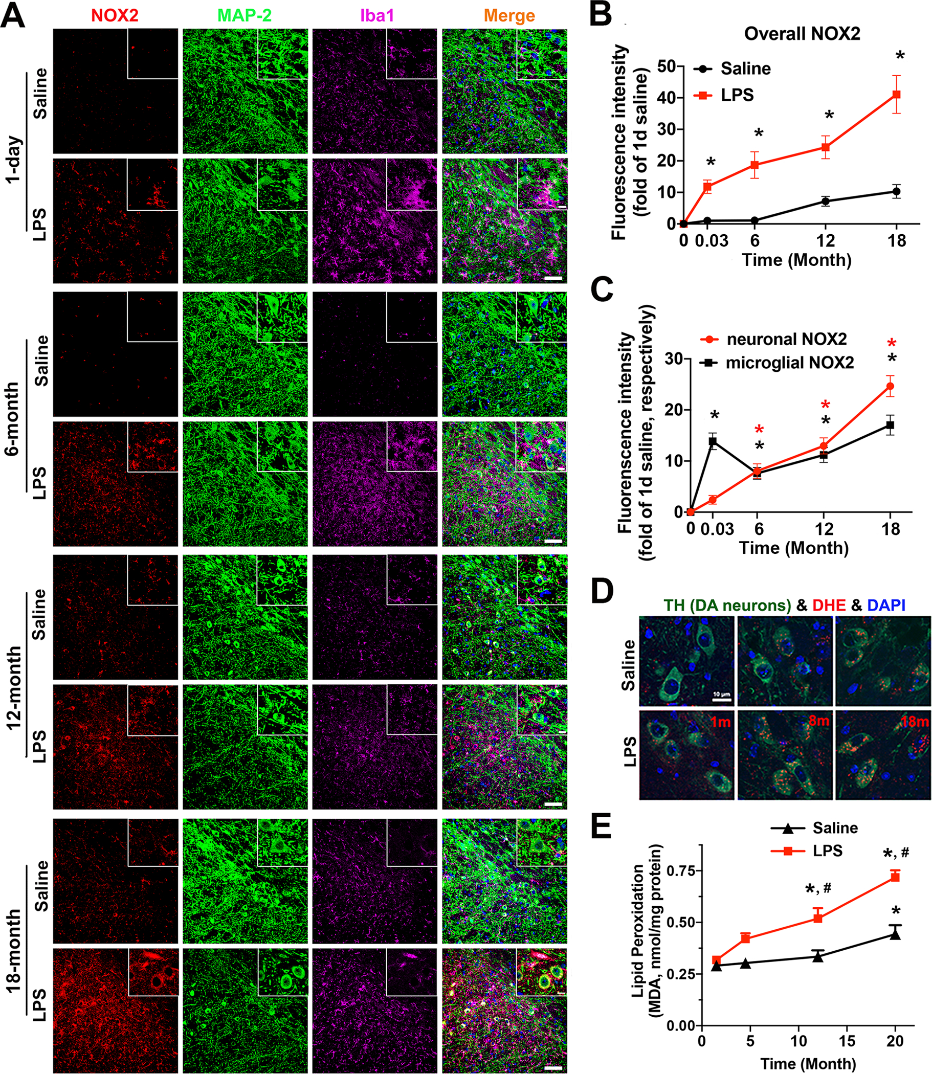
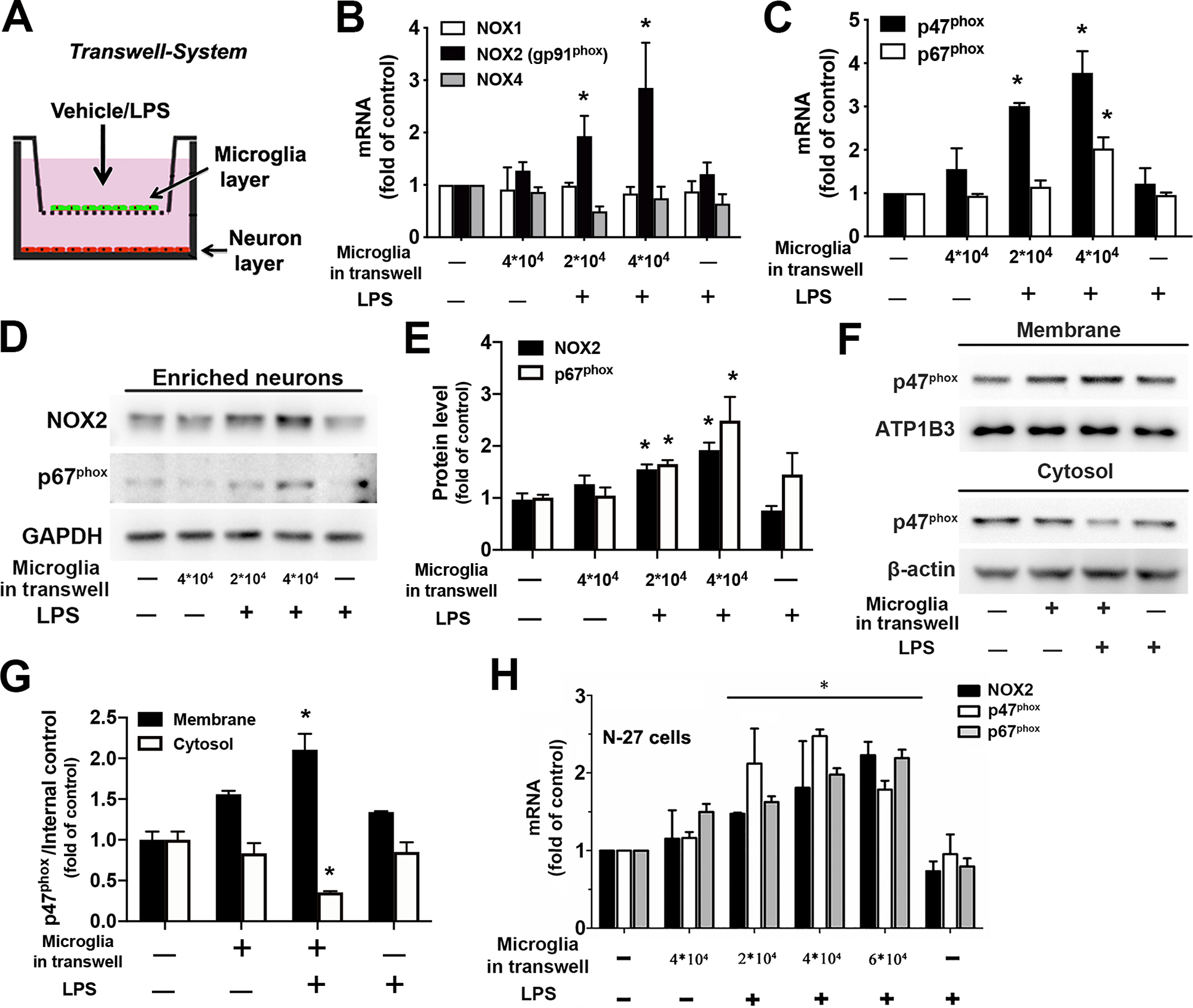
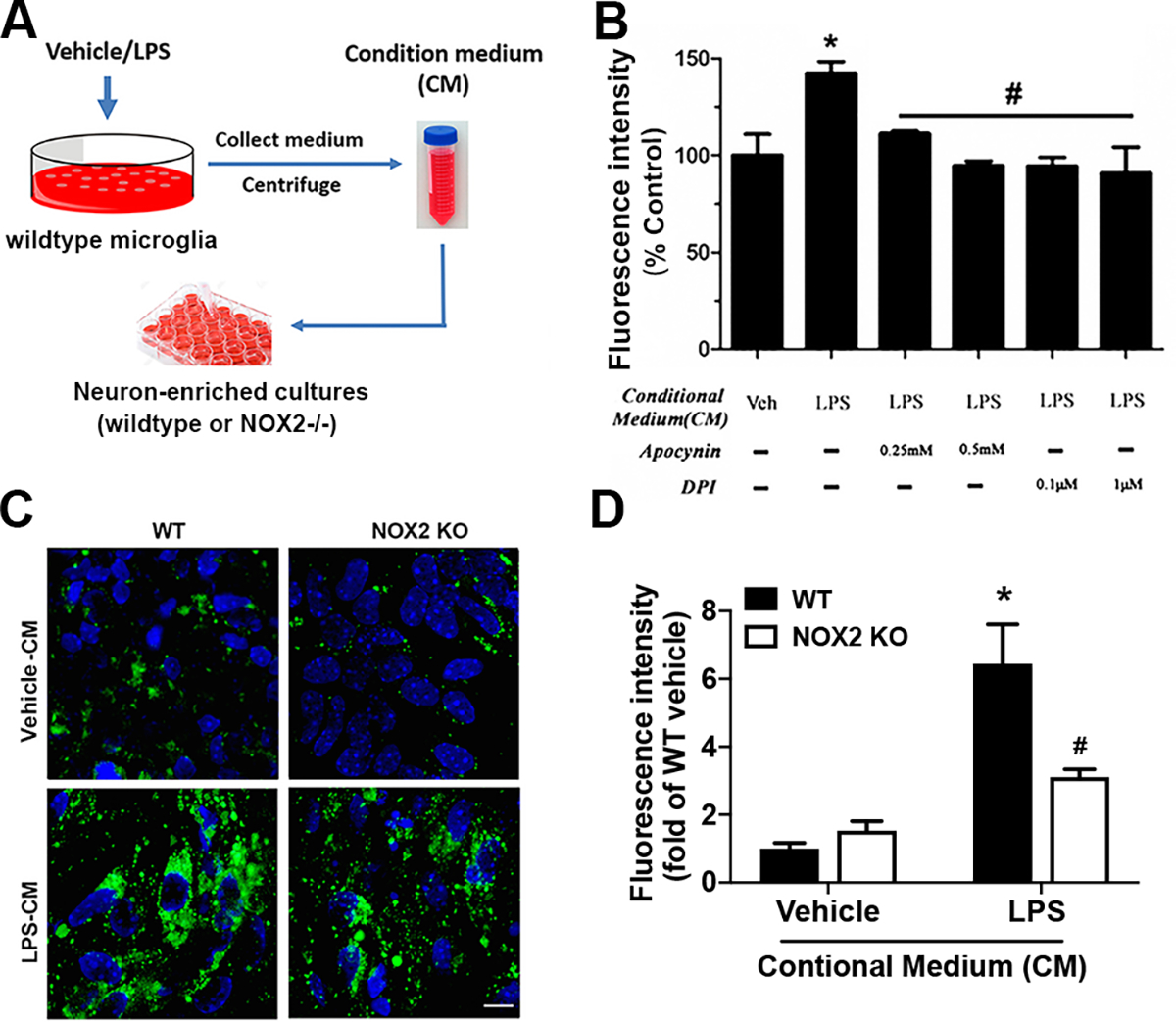
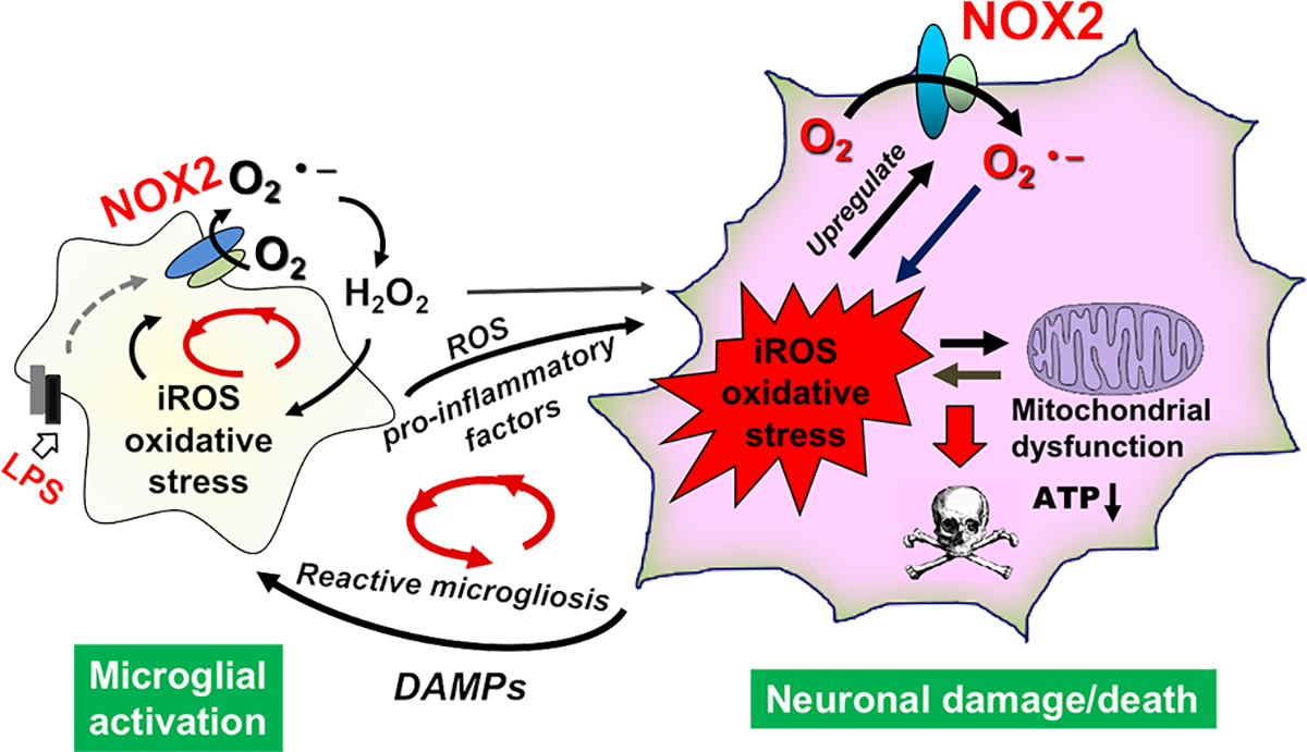
Strong evidence indicates critical roles of NADPH oxidase (a key superoxide-producing enzyme complex during inflammation) in activated microglia for mediating neuroinflammation and neurodegeneration. However, little is known about roles of neuronal NADPH oxidase in neurodegenerative diseases. This study aimed to investigate expression patterns, regulatory mechanisms and pathological roles of neuronal NADPH oxidase in inflammation-associated neurodegeneration. The results showed persistent upregulation of NOX2 (gp91phox; the catalytic subunit of NADPH oxidase) in both microglia and neurons in a chronic mouse model of Parkinson's disease (PD) with intraperitoneal LPS injection and LPS-treated midbrain neuron-glia cultures (a cellular model of PD). Notably, NOX2 was found for the first time to exhibit a progressive and persistent upregulation in neurons during chronic neuroinflammation. While primary neurons and N27 neuronal cells displayed basal expression of NOX1, NOX2 and NOX4, significant upregulation only occurred in NOX2 but not NOX1 or NOX4 under inflammatory conditions. Persistent NOX2 upregulation was associated with functional outcomes of oxidative stress including increased ROS production and lipid peroxidation. Neuronal NOX2 activation displayed membrane translocation of cytosolic p47phox subunit and was inhibited by apocynin and diphenyleneiodonium chloride (two widely-used NADPH oxidase inhibitors). Importantly, neuronal ROS production, mitochondrial dysfunction and degeneration induced by inflammatory mediators in microglia-derived conditional medium were blocked by pharmacological inhibition of neuronal NOX2. Furthermore, specific deletion of neuronal NOX2 prevented LPS-elicited dopaminergic neurodegeneration in neuron-microglia co-cultures separately grown in the transwell system. The attenuation of inflammation-elicited upregulation of NOX2 in neuron-enriched and neuron-glia cultures by ROS scavenger N-acetylcysteine indicated a positive feedback mechanism between excessive ROS production and NOX2 upregulation. Collectively, our findings uncovered crucial contribution of neuronal NOX2 upregulation and activation to chronic neuroinflammation and inflammation-related neurodegeneration. This study reinforced the importance of developing NADPH oxidase-targeting therapeutics for neurodegenerative diseases.

Keywords: Microglia; Mitochondrial dysfunction; NOX2; Neurodegeneration; Neuronal NADPH oxidase; Oxidative stress.


Conflict of interest statement

Declaration of competing interest We state ‘Declarations of interest: none’ in the title page and have a Section “Competing interests” and statement “The authors declare that they have no competing interests” in the manuscript.

Figures

Fig. 1
Fig. 1. LPS induced persistent upregulation of NOX2 in both microglia and neurons in neuron-glia cultures.
A-C Rat midbrain neuron-glia cultures were challenged with vehicle or LPS (10 ng/ml; the concentration used in all in vitro experiments unless otherwise indicated). The expression of mRNA and protein of NOX2 (gp91phox) was measured by using RT-PCR (A) and Western blotting (B) with densitometry analysis (C) at indicated time points. Results are mean ± SEM of three experiments performed in triplicate (n = 3). *p < 0.05 compared with time-matched vehicle-treated controls. D, E Rat midbrain neuron-glia cultures were treated with vehicle or LPS for 6 days. D Representative immunohistochemical analysis showed that NOX2 was persistently upregulated during chronic neuroinflammation. E Representative images of double-labelled immunofluorescence showed increased expression of NOX2 (green) in neurons (red staining for neuronal maker Nissl) after LPS treatment. All immunoblotting and immunostaining images were representative of three independent experiments. Scale bars in (D) and (E) are 50 and 10 μm, respectively.
Fig. 2
Fig. 2. The SN of a chronic mouse model of PD showed persistent upregulation of NOX2 in microglia and neurons as well as elevated oxidative stress and lipid peroxidation.
A-C Frozen brain slices (16 μm in thickness) from C57BL/6 mice with an intraperitoneal injection of saline or LPS (5 mg/kg; 15 × 106 EU/kg; the dose used for all in vivo experiments) were triple-stained for NOX2 (red), MAP-2 (green), and Iba-1 (purple) at 1 day or 6, 12 and 18 months after the injection. A The representative confocal fluorescence images of the SN region were shown. The scale bar is 50 μm. The zoomed images were shown in the upper right corner of the corresponding images, and the scale bar in the zoomed images is 10 μm. B The quantification of fluorescence intensity of overall levels of NOX2 in the SN of saline- and LPS-injected mice. C The quantification of fluorescence intensity of microglial NOX2 and neuronal NOX2 at the indicated time points after vehicle/LPS injection. *p < 0.05 versus saline-injected mice. D At 1, 8, and 18 months after LPS/saline injection, mice were injected intraperitoneally with DHE (30 mg/kg). Brains were then harvested 30 minutes later. Fluorescent images of mouse brain cryosections showed double-labeling for ethidium and 2-hydroxyethidium (red) and TH (green) after DHE oxidation. E Mice injected with saline or LPS were scarified at 1.5, 4.5, 12, and 20 months after the injection. Lipid peroxidation was evaluated by the measurement of the level of MDA. Data are mean ± SEM from 5 mice for each time point (B, C and E). *p < 0.05 versus the corresponding 1.5-month group. #p < 0.05 versus time matched saline-injected groups. All immunostaining images were representative of 5 mice for each time point (A, D).
Fig. 3
Fig. 3. Upregulation and activation of NDAPH oxidase in neurons under inflammatory conditions.
A-G. Rat neurons and microglia were separately cultured in a transwell system. This was achieved by seeding different amount of enriched microglia on the porous membrane of transwell inserts that were placed above neuronal layer grown in the well of the plate. At 24 hours after LPS or vehicle treatment of the co-cultures, neurons were collected for the measurement of mRNA or protein of subunits of NADPH oxidase. A Schematic diagram of the transwell cell culture system. B The mRNA expression of the catalytic subunit of NADPH oxidase, NOX1, NOX2 (gp91phox) and NOX4 isoforms in neurons was determined by using RT-PCR. C RT-PCR detected mRNA expression of cytosolic subunits (p47phox and p67phox) in neurons. D The protein level of NOX2 and p67phox in neurons was determined by Western blot. E The ratios of the densitometry values of NOX2 or p67phox relative to GAPDH in (C) were normalized to vehicle-treated control. F Membrane and cytosolic fractions were isolated from neurons and used for examination of membrane translocation of cytosolic subunit p47phox by Western blot. ATP1B3 and β-actin were used to monitor loading errors of membrane and cytosolic proteins respectively. G The densitometry values of p47phox relative to ATP1B3 or β-actin in (F) were normalized to vehicle-treated control. H The mRNA expression of major subunits of NADPH oxidase (NOX2, p47phox and p67phox) in N27 neuronal cell line was examined by RT-PCR after N27-microglia co-cultures grown in the transwell system were treated with LPS/vehicle for 24 hours. Data are shown as means ± SEM from three independent experiments performed in triplicate (B, C, E, G and H; n = 3). *p < 0.05 compared with the corresponding saline-treated controls.
Fig. 4
Fig. 4. Inflammation-induced neuronal NOX2-dependent ROS production in neurons.
A Schematic diagram of the collection of conditional medium (CM) from vehicle-/LPS-treated microglia-enriched cultures and the treatment of neuron-enriched cultures with the CM. Mouse microglia (106 cells/well) were treated with vehicle or LPS for 24 hours. Supernatants were collected and centrifuged to remove cells and/or cell debris. One milliliter of CM was added to each well containing neuron-enriched cultures. B Measurement of iROS-dependent intracellular H2DCFDA oxidation using fluorescence microplate reader to detect iROS in midbrain neuron-enriched cultures that were pretreated with apocynin (0.25 or 0.5 mM) or DPI (0.1 or 1 μM) for 30 minutes and then incubated with the CM for 24 hours. C, D Midbrain neuron-enriched cultures prepared from wildtype or NOX2−/− mice were incubated for 24 hours with the CM. Representative confocal images of a fluorescent adduct of CM-H2DCFDA after its iROS-dependent oxidation (C) and quantification of the fluorescence intensity by Image J (D). Data are mean ± SEM from three independent experiments performed in triplicate (B, D; n = 3). *p < 0.05 compared with the corresponding control cultures incubated with CM from vehicle-treated wildtype microglia. #p < 0.05 compared with the corresponding wildtype neuronal cultures incubated with CM from LPS-treated wildtype microglia (B, D). The scale bar is 20 μm.
Fig. 5
Fig. 5. A positive feedback mechanism underlies the sustained NOX2 upregulation.
A Rat midbrain neuron-enriched cultures were treated with vehicle or NAC (a commonly used ROS scavenger; 5 mM) and incubated for 24 hours with the CM from vehicle-/LPS-treated microglia-enriched cultures. The mRNA expression of NOX2 in neurons was determined by using RT-PCR. B-D Rat midbrain neuron-glia cultures were challenged with LPS with or without pretreatment or post-treatment with NAC (5 mM) at 30 min before or 1 day after LPS challenge. The expression of NOX2 mRNA and protein was determined using RT-PCR (B) and Immunoblotting (C, D). Pre- and post-treatment with NAC resulted in a significant reduction in the level of NOX2 mRNA and protein, indicating inflammation-associated ROS production upregulated NOX2 expression. Data are mean ± SEM from three independent experiments in triplicate (n = 3). *p < 0.05 and #p < 0.05 compared with neuron-enriched cultures incubated with CM from vehicle-treated and LPS-treated microglia respectively in (A). *p < 0.05 compared with vehicle-treated neuron-glia cultures in (B, D). #p < 0.05 compared with LPS-treated neuron-glia cultures (B, D). C: control; L: LPS; N: NAC.
Fig. 6
Fig. 6. Neuronal NOX2 activation triggered by inflammatory factors induced neuronal mitochondrial dysfunction and dopaminergic neurotoxicity.
A, B Intracellular ATP (A) and mitochondrial membrane potential (B) were measured in midbrain neuron-enriched cultures that were pretreated with apocynin (0.5 mM) for 30 minutes and then incubated with CM from vehicle- or LPS-treated microglia. C Midbrain neuron-enriched cultures were pretreated with apocynin (0.25 or 0.5 mM) for 30 minutes and then incubated with the CM from vehicle- or LPS-treated microglia. [3H]dopamine uptake assay was preformed to detect dopaminergic neurodegeneration 7 days after the addition of CM. D, E Midbrain neuron-enriched cultures prepared from wildtype or NOX2−/− mice with or without co-culture with wildtype microglia and vehicle/LPS treatment. Neurodegeneration was then determined by [3H]dopamine uptake assay (D) and visualized after immunostaining for TH (E) at 7 days after the treatment. Data are mean ± SEM from three independent experiments performed in triplicate (A-D; n = 3). *p < 0.05 and #p < 0.05 compared with the corresponding neuronal cultures incubated with CM from vehicle-treated and LPS-treated wildtype microglia respectively (A-C). *p < 0.05 compared with the wildtype neurons co-cultured with wildtype microglia and treated with vehicle (D). #p < 0.05 compared with the wildtype neurons co-cultured with wildtype microglia and treated with the same concentration of LPS (D). The images presented in (E) are representative of three independent experiments. The scale bar is 5 μm.
Fig. 7
Fig. 7. Multiple positive feed-back cycles sustained chronic neuroinflammation and drove progressive neurodegeneration.
Initially, LPS triggered the activation of microglial NADPH oxidase and generated a large amount of superoxide radical, which was converted to H2O2 through superoxide dismutase. Cell membrane permeable H2O2 then entered nearby neurons to increase neuronal iROS; meanwhile pro-inflammatory factors released from activated microglia acted on neurons. As a result, expression of NOX2 was upregulated in neurons, and more neuronal NOX2-dependent iROS were produced in neurons. These results indicated the formation of a positive feedback between ROS production and neuronal NOX2 upregulation. Excessive iROS caused mitochondrial damages and dysfunction leading to leak of superoxide. Therefore, a “ROS-producing-ROS” vicious cycle was formed within neurons inducing persistent oxidative damages. As neuroinflammation and oxidative stress continued, the vicious positive feedback loop within neurons caused neuronal damages/death and leak of danger-associated molecular patterns (DAMPs) to further trigger the reactive microgliosis and to form another positive feedback cycle between dysregulated microglial activation and neuronal damages. There were similar positive feedback cycles between ROS production and NOX2 upregulation/activation or between NOX2-derived ROS and mitochondria-derived ROS in microglia. O2.−: superoxide radical; H2O2: hydrogen peroxide.

References

    1. Block ML, Zecca L, Hong JS, Microglia-mediated neurotoxicity: uncovering the molecular mechanisms, Nat Rev Neurosci 8(1) (2007) 57–69. - PubMed
    1. Simpson DS, Oliver PL, ROS generation in microglia: understanding oxidative stress and inflammation in neurodegenerative disease, Antioxidants 9(8) (2020) 743. - PMC - PubMed
    1. Tu D, Gao Y, Yang R, Guan T, Hong JS, Gao HM, The pentose phosphate pathway regulates chronic neuroinflammation and dopaminergic neurodegeneration, J Neuroinflammation 16(1) (2019) 1–17. - PMC - PubMed
    1. Geng L, Fan LM, Liu F, Smith C, Li JM, Nox2 dependent redox-regulation of microglial response to amyloid-β stimulation and microgliosis in aging, Sci Rep 10(1) (2020) 1–11. - PMC - PubMed
    1. Moehle MS, West AB, M1 and M2 immune activation in Parkinson’s Disease: Foe and ally?, Neuroscience 302 (2015) 59–73. - PMC - PubMed