. 2023 Apr:28:115-123.
doi: 10.1016/j.jtos.2023.02.003. Epub 2023 Mar 3.

Genipin increases extracellular matrix synthesis preventing corneal perforation

Affiliations

Genipin increases extracellular matrix synthesis preventing corneal perforation

Christopher Donovan et al. Ocul Surf. 2023 Apr.

Abstract

Purpose: Corneal melting and perforation are feared sight-threatening complications of infections, autoimmune disease, and severe burns. Assess the use of genipin in treating stromal melt.

Methods: A model for corneal wound healing was created through epithelial debridement and mechanical burring to injure the corneal stromal matrix in adult mice. Murine corneas were then treated with varying concentrations of genipin, a natural occurring crosslinking agent, to investigate the effects that matrix crosslinking using genipin has in wound healing and scar formation. Genipin was used in patients with active corneal melting.

Results: Corneas treated with higher concentrations of genipin were found to develop denser stromal scarring in a mouse model. In human corneas, genipin promoted stromal synthesis and prevention of continuous melt. Genipin mechanisms of action create a favorable environment for upregulation of matrix synthesis and corneal scarring.

Conclusion: Our data suggest that genipin increases matrix synthesis and inhibits the activation of latent transforming growth factor-β. These findings are translated to patients with severe corneal melting.

Keywords: Collagens; Fibroblasts; Stroma; Wound.


Conflict of interest statement

Declaration of competing interest None of the authors has any commercial affiliations or consultancies, stock or equity interests, or patent-licensing arrangements that could be considered to pose a financial conflict of interest related to this manuscript. EME was a consultant for GSK in a matter not related to what is investigated in this manuscript.

Figures

Figure 1.
Figure 1.. Genipin exposure increases corneal scarring at 3 weeks only if stromal injury is performed.
(A,B) No significant haze is noticeable after mild stromal injury without topical genipin application or epithelial debridement and exposure to genipin without stromal injury. (C, D, E and F) Increasing corneal haze and scarring noted with higher genipin concentrations 0.1%(C) , 0.125 % (D) or 0.25% (E) for 2 mins or 0.5% for 5 mins (F). Increasing dose or exposure time resulted in increased haze and scar as graded using a modified Fantes scale. **P < 0.01. NS: not significant.
Figure 2.
Figure 2.. Increased cellular synthetic activity and deposition of a disorganized matrix following genipin treatment.
Topical application of 2.5 mg/ml genipin for 5 minutes in vehicle (HBSS) following injury vs vehicle only for 5 mins. (A1) Denser scarring and haze are observed in eyes treated with genipin following mechanical burr injury. Collagen IV and fibronectin expression was upregulated. In contrast, in eyes treated with vehicle only following injury, Collagen IV and fibronectin expression was minimal (A2). Second harmonic generation imaging shows increased cellularity, higher forward scattering signaling suggesting a more disorganized deposition of collagen fibrils following injury when genipin was used (B1) compared to vehicle (B2). Increased expression of PAI-1, collagen I and α-smooth muscle actin in stromal matrix after genipin use. Negative controls showed no reactivity. Nuclei were stained with DAPI (blue). At least 3 samples were used. *P < 0.05; **P < 0.01. Bar 50μm.
Figure 3.
Figure 3.. Keratocyte derived cell lineage responds to matrix stiffness by regulating cell morphology and function.
Our I-KeramTmG mouse strain facilitates isolation of keratocyte derived cell lineages expressing eGFP. (A) In this corneal histology section, epithelium and endothelium express dTm while most but not all stromal cells express keratocan, see asterisks (eGFP). (B) Increasing matrix stiffness regulates keratocyte derived cells morphology. Broader cells noted with increasing matrix stiffness. (C) Increasing matrix stiffness activates TGF-β signaling as suggested by increased PAI-1 mRNA in keratocyte derived fibroblasts. Collagen I mRNA synthesis is upregulated by increased matrix stiffness in keratocyte derived fibroblasts. White bar represents mRNA obtained from expanded cells on 2kPa and black bars from cells expanded on 64 kPa. Nuclei were stained with DAPI (blue). At least 3 samples were used. *P < 0.05; **P < 0.01. Bar 50μm.
Figure 4.
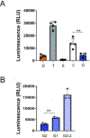
Figure 4.. Stromal crosslinking and stiffening impair human corneal stromal TGF-β activation.
(A) Luciferase assay was used to functionally quantify TGF-β availability in human corneal stromas crosslinked with genipin. Stromal discs, 2mm in diameter after epithelial removal, were crosslinked in a solution of 2 mg/ml of genipin for 30 minutes (G) or placed in HBSS (vehicle control, V). Tissue was then washed out 3 times and heated at 80°C for 10 mins to activate latent TGF-β. D denotes basal levels of TGF-β by culturing reporter cells only in DMEM, T denotes reporter cells stimulated by 10 ng/ml human recombinant TGF-β1. A TGF-β specific blocker, SB431542 (S) demonstrate that reporter cells were responding to TGF-β signaling and no other cytokines. ** p=0.005; (B) Dose response experiment using three different genipin concentrations: G2 (2 mg/ml). G1 (1 mg/ml) and G0.2 (0.2 mg/ml). RLU: relative light unit. **p=0.0019. Experiments were repeated 4 times to ensure reproducibility.
Figure 5.
Figure 5.. Clinical case demonstrates genipin effects in case of active ulceration.
Illustrative case showing a red eye with progressive stromal thinning mostly located in the corneal periphery and midperiphery despite continuous broad spectrum antibiotic treatment (A). Eye appears quiet and stromal thinning improved 1 week after genipin application to affected area. Note light stromal blue hue (B). Quiet eye with peripheral vascularization and haze at no risk of perforation. Slight blue hue still present. This case represents patient number three in Table 1.
Figure 6.
Figure 6.
Graphical summary of mechanism of action for genipin.

References

    1. Espana EM, Birk DE. Composition, structure and function of the corneal stroma. Exp Eye Res. 2020:108137. - PMC - PubMed
    1. Hassell JR, Birk DE. The molecular basis of corneal transparency. Exp Eye Res. 2010;91:326–35. - PMC - PubMed
    1. Maurice DM. The structure and transparency of the cornea. J Physiol. 1957;136:263–86. - PMC - PubMed
    1. Meek KM, Knupp C. Corneal structure and transparency. Prog Retin Eye Res. 2015;49:1–16. - PMC - PubMed
    1. Jabbehdari S, Rafii AB, Yazdanpanah G, Hamrah P, Holland EJ, Djalilian AR. Update on the Management of High-Risk Penetrating Keratoplasty. Curr Ophthalmol Rep. 2017;5:38–48. - PMC - PubMed

Publication types