Effect of commercially pure titanium implant coated with calcium carbonate and nanohydroxyapatite mixture on osseointegration
- PMID: 36873118
- PMCID: PMC9979178
- DOI: 10.25122/jml-2022-0049
Effect of commercially pure titanium implant coated with calcium carbonate and nanohydroxyapatite mixture on osseointegration
Abstract
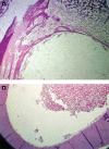
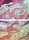
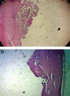
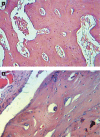
In this research, rabbit femurs were implanted with CP Ti screws coated with a combination of CaCO3 and nanohydroxyapatite, and the effect on osseointegration was assessed using histological and histomorphometric examination at 2 and 6 weeks. CaCO3 and nanohydroxyapatite were combined with the EPD to coat the surfaces of the CP Ti screws. The femurs of five male rabbits were implanted with coated and uncoated implant screws. Healing time was divided into two groups (2 and 6 weeks). After 2 and 6 weeks of implantation, the histological examination revealed an increase in the growth of bone cells for coated screws, and the histomorphometric analysis revealed an increase in the percentage of new bone formation (after 6 weeks, 5.08% for coated implants and 3.66% for uncoated implants). In addition, the uncoated implant, the CP Ti implant coated with a combination of CaCO3 and nanohydroxyapatite, stimulated early bone development after two weeks and mineralization and maturation after six weeks.
Keywords: calcium carbonate; electrophoretic; histomorphometric; nanohydroxyapatite; osseointegration.
©2022 JOURNAL of MEDICINE and LIFE.
Conflict of interest statement
The authors declare no conflict of interest.
Figures


Similar articles
-
Effects of magnesium-substituted nanohydroxyapatite coating on implant osseointegration.Clin Oral Implants Res. 2013 Aug;24 Suppl A100:34-41. doi: 10.1111/j.1600-0501.2011.02362.x. Epub 2011 Dec 6. Clin Oral Implants Res. 2013. PMID: 22145854
-
Enhanced biocompatibility and osseointegration of calcium titanate coating on titanium screws in rabbit femur.J Huazhong Univ Sci Technolog Med Sci. 2017 Jun;37(3):362-370. doi: 10.1007/s11596-017-1741-9. Epub 2017 Jun 6. J Huazhong Univ Sci Technolog Med Sci. 2017. PMID: 28585129
-
The correlation between osseointegration and bonding strength at the bone-implant interface: In-vivo & ex-vivo investigations on hydroxyapatite and hydroxyapatite/titanium coatings.J Biomech. 2022 Nov;144:111310. doi: 10.1016/j.jbiomech.2022.111310. Epub 2022 Sep 19. J Biomech. 2022. PMID: 36162145
-
Osseointegration is improved by coating titanium implants with a nanostructured thin film with titanium carbide and titanium oxides clustered around graphitic carbon.Mater Sci Eng C Mater Biol Appl. 2017 Jan 1;70(Pt 1):264-271. doi: 10.1016/j.msec.2016.08.076. Epub 2016 Aug 31. Mater Sci Eng C Mater Biol Appl. 2017. PMID: 27770890
-
Review of PEEK implants and biomechanical and immunological responses to a zirconium phosphate nano-coated PEEK, a blasted PEEK, and a turned titanium implant surface.Am J Dent. 2022 Jun;35(2):152-160. Am J Dent. 2022. PMID: 35798711 Review.
Cited by
-
Biomimetic Scaffolds of Calcium-Based Materials for Bone Regeneration.Biomimetics (Basel). 2024 Aug 24;9(9):511. doi: 10.3390/biomimetics9090511. Biomimetics (Basel). 2024. PMID: 39329533 Free PMC article. Review.
-
Exploring the potential of a newly developed pectin-chitosan polyelectrolyte composite on the surface of commercially pure titanium for dental implants.Sci Rep. 2023 Dec 14;13(1):22203. doi: 10.1038/s41598-023-48863-2. Sci Rep. 2023. PMID: 38097618 Free PMC article.
-
Citrus-Fruit-Based Hydroxyapatite Anodization Coatings on Titanium Implants.Materials (Basel). 2025 Mar 5;18(5):1163. doi: 10.3390/ma18051163. Materials (Basel). 2025. PMID: 40077388 Free PMC article.
References
-
- Brunski J, Glantz PO, Helms JA, Nanci A. Transfer of mechanical local across the interface. Osseointegration Book. In: PI Branemark., editor. Berlin: Quintessenz; 2005. pp. 209–249.
-
- Mavrogenis AF, Dimitriou R, Parvizi J, Babis GC. Biology of implant osseointegration. J Musculoskelet Neuronal Interact. 2009;9(2):61–71. - PubMed
MeSH terms
Substances
LinkOut - more resources
Full Text Sources
Miscellaneous
