Case Reports
. 2022 Oct 5;9(1):20220046.
doi: 10.1259/bjrcr.20220046. eCollection 2023 Feb.

Gout of the temporomandibular joint and review of the literature

Affiliations
Case Reports

Gout of the temporomandibular joint and review of the literature

Joel Hng et al. BJR Case Rep. .

Abstract

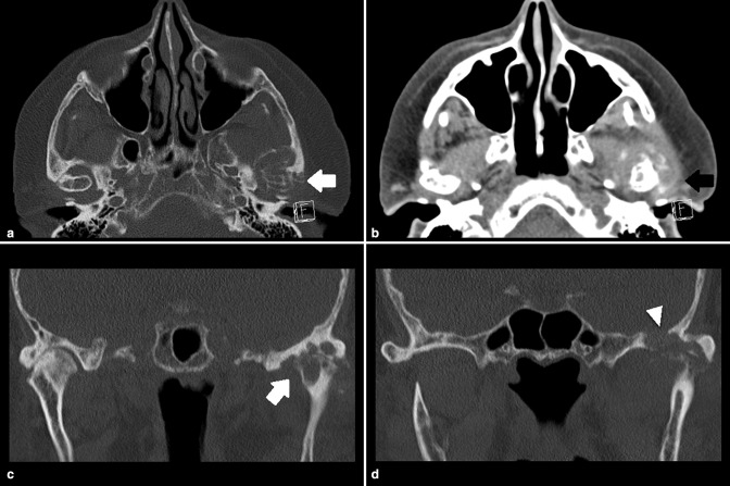
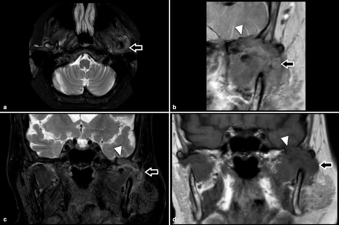
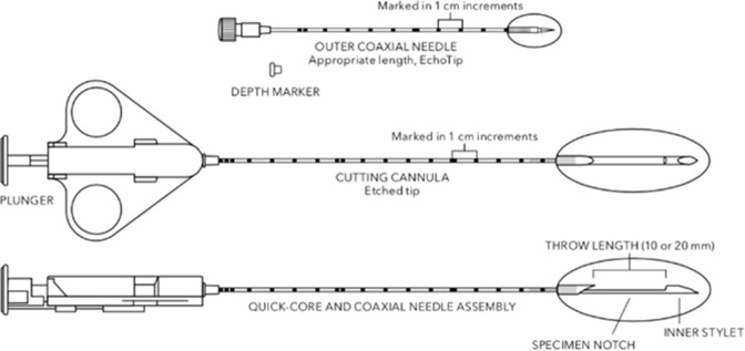
Gout is a disease characterised by abnormal deposition of monosodium urate crystals, typically affecting the extremities. This report describes a rare case of gout affecting the left temporomandibular joint with erosion of the skull base. A diagnosis of gout was suspected based on CT and MRI and confirmed with CT-guided biopsy. The temporomandibular joint is an uncommon location for a first presentation of gout, with very few cases documented and only three cases of skull base involvement reported in the English literature previously. Given its radiological appearance, it can easily be misdiagnosed as other erosive arthropathies or malignancy. Our paper highlights an unusual location for the first and only manifestation of gout and offers some diagnostic and treatment ideas that may help clinicians to identify and manage this disease.


Figures

Figure 1.
Figure 1.
CT images showing relatively well-defined left TMJ articular and juxtaarticular erosions (white arrows) with through and through defect of the skull base (arrow head) and surrounding dense soft tissue thickening (black arrow). TMJ, temporomandibular joint.
Figure 2.
Figure 2.
Axial and Coronal fat suppressed T2 (a and c), targeted coronal PD (b) and coronal T1 (d) MRI images showing erosions with through and through defect of the skull base (arrow head) and surrounding lobulated soft tissue thickening (arrows). PD, proton density.
Figure 3.
Figure 3.
Quick-Core Biopsy Needle set, containing a Quick-Core needle and a coaxial outer needle. The specimen notch can be opened by retracting the cutting cannula to 10 mm or 20 mm in length, depending on the size of the lesion.

Similar articles

Cited by

References

    1. Oliveira INF, Gomes RCF, Dos Santos RR, Oliveira T de P, Pereira LLC, Mainenti P. Gout of the temporomandibular joint: report of a case. Int Arch Otorhinolaryngol 2014; 18: 316–18. doi: 10.1055/s-0033-1363464 - DOI - PMC - PubMed
    1. Suba Z, Takács D, Gyulai-Gaál S, Fancsaly AJ, Szabó G, Undt G, et al. . Tophaceous gout of the temporomandibular joint: a report of 2 cases. J Oral Maxillofac Surg 2009; 67: 1526–30. doi: 10.1016/j.joms.2008.12.019 - DOI - PubMed
    1. Barthélémy I, Karanas Y, Sannajust J-P, Emering C, Mondié J-M. Gout of the temporomandibular joint: pitfalls in diagnosis. J Craniomaxillofac Surg 2001; 29: 307–10. doi: 10.1054/jcms.2001.0244 - DOI - PubMed
    1. Bhattacharyya I, Chehal H, Gremillion H, Nair M, Odont L. Gout of the temporomandibular joint: a review of the literature. J Am Dent Assoc 2010; 141: 979–85. doi: 10.14219/jada.archive.2010.0311 - DOI - PubMed
    1. Sidari A, Hill E. Diagnosis and treatment of gout and pseudogout for everyday practice. Prim Care 2018; 45: 213–36. doi: 10.1016/j.pop.2018.02.004 - DOI - PubMed

Publication types