. 2023 Feb 28:11:tkac055.
doi: 10.1093/burnst/tkac055. eCollection 2023.

Nuclear fragile X mental retardation-interacting protein 1-mediated ribophagy protects T lymphocytes against apoptosis in sepsis

Affiliations

Nuclear fragile X mental retardation-interacting protein 1-mediated ribophagy protects T lymphocytes against apoptosis in sepsis

Peng-Yue Zhao et al. Burns Trauma. .

Abstract

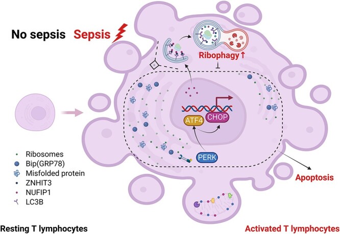
Background: Ribophagy is a selective autophagic process that specifically degrades dysfunctional or superfluous ribosomes to maintain cellular homeostasis. Whether ribophagy can ameliorate the immunosuppression in sepsis similar to endoplasmic reticulum autophagy (ERphagy) and mitophagy remains unclear. This study was conducted to investigate the activity and regulation of ribophagy in sepsis and to further explore the potential mechanism underlying the involvement of ribophagy in T-lymphocyte apoptosis.

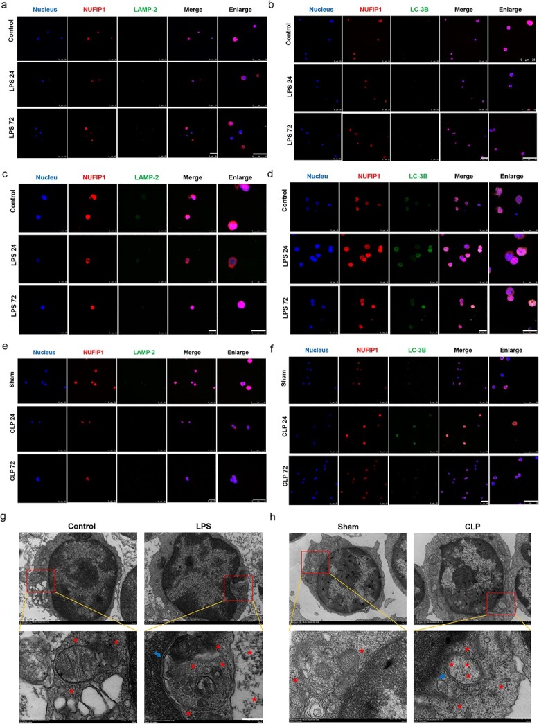
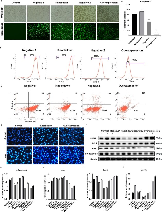
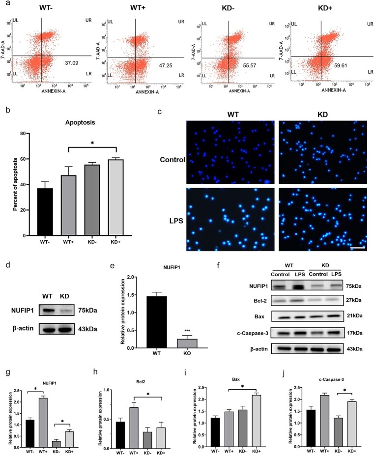
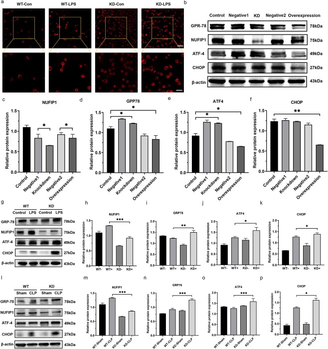
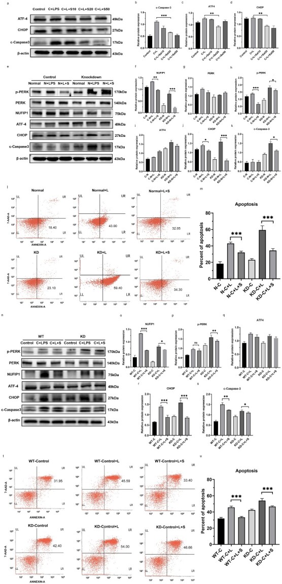
Methods: The activity and regulation of nuclear fragile X mental retardation-interacting protein 1 (NUFIP1)-mediated ribophagy in T lymphocytes during sepsis were first investigated by western blotting, laser confocal microscopy and transmission electron microscopy. Then, we constructed lentivirally transfected cells and gene-defective mouse models to observe the impact of NUFIP1 deletion on T-lymphocyte apoptosis and finally explored the signaling pathway associated with T-cell mediated immune response following septic challenge.

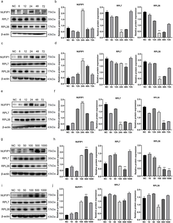
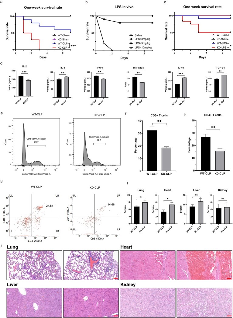
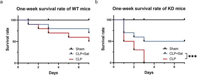
Results: Both cecal ligation and perforation-induced sepsis and lipopolysaccharide stimulation significantly induced the occurrence of ribophagy, which peaked at 24 h. When NUFIP1 was knocked down, T-lymphocyte apoptosis was noticeably increased. Conversely, the overexpression of NUFIP1 exerted a significant protective impact on T-lymphocyte apoptosis. Consistently, the apoptosis and immunosuppression of T lymphocytes and 1-week mortality rate in NUFIP1 gene-deficient mice were significantly increased compared with those in wild-type mice. In addition, the protective effect of NUFIP1-mediated ribophagy on T lymphocytes was identified to be closely related to the endoplasmic reticulum stress apoptosis pathway, and PERK-ATF4-CHOP signaling was obviously involved in downregulating T-lymphocyte apoptosis in the setting of sepsis.

Conclusions: NUFIP1-mediated ribophagy can be significantly activated to alleviate T lymphocyte apoptosis through the PERK-ATF4-CHOP pathway in the context of sepsis. Thus, targeting NUFIP1-mediated ribophagy might be of importance in reversing the immunosuppression associated with septic complications.

Keywords: Apoptosis; Autophagy; Immunosuppression; Lipopolysaccharide; Lymphocyte; Mitophagy; NUFIP1; Ribophagy; Sepsis.


Figures

Figure 1
Figure 1
Time-effect and dose-effect responses of ribophagy in sepsis. (a, b) The time-effect response of splenic CD4+ T lymphocytes in vitro. After LPS stimulation for 6, 12, 24, 48 and 72 h, the protein levels of NUFIP-1 in splenic CD4+ T lymphocytes showed a trend of an initial increase and then decrease, and they peaked at 24 h. However, RPL7 and RPL26 expression exhibited a trend of an initial decrease and then increase, with the lowest value observed at 24 h. (c, d) Time-effect response of Jurkat cells. (e, f) The in vivo time-effect response. (gj) The in vitro dose-effect experiment. After stimulating splenic CD4+ T lymphocytes or Jurkat cells with 10, 50, 100, 500 and 1000 ng/ml LPS for 24 h, expression of the NUFIP1 protein was upregulated in the stimulated group compared to the blank control group, while that of RPL7 and RPL26 was downregulated. Among the groups, the 500 ng/ml LPS stimulation group exhibited the most significant increase or decrease. One-way ANOVA was applied to test the statistical significance. Data are expressed as means ± SEM; **p < 0.01, ***p < 0.001. NC normal control group, NUFIP1 nuclear fragile X mental retardation-interacting protein 1, RPL7 ribosomal protein L7, RPL26 ribosomal protein L26, LPS lipopolysaccharide, ANOVA analysis of variance, SEM standard error of mean
Figure 2
Figure 2
Ribophagy was significantly activated in sepsis. (a, b) LSCM examination indicated that the NUFIP-1 protein was scattered in both the cytoplasm and nucleus but mainly located in the nucleus. The expression of NUFIP1 was significantly increased in splenic CD4+ T lymphocytes after 500 ng/ml LPS stimulation for 24 h compared with control treatment, and NUFIP1 shuttling from the nucleus to the cytoplasm was observed. In addition, the colocalization of NUFIP1 with LC-3B and LAMP-2 was substantially intensified in the LPS-stimulated group in comparison to the control group. When the LPS stimulation time was increased to 72 h, the above changes gradually decreased, becoming close to the level observed before LPS stimulation (scale bar: 25 μm). (c, d) The expression of NUFIP1 and the fusion of NUFIP1 with LC-3B and LAMP-2 in Jurkat cells after 500 ng/ml LPS stimulation for 24 and 72 h compared with control groups (scale bar: 25 μm). (e, f) The expression of NUFIP1 and the fusion of NUFIP1 with LC-3B and LAMP-2 in splenic CD4+ T lymphocytes after CLP for 24 and 72 h compared with that in the sham group (scale bar: 25 μm). (g, h) TEM showed that compared with the control or sham group, the LPS or CLP group showed an obviously swollen ER in splenic CD4+ T lymphocytes and autophagosomes containing a large number of ribosomes to be degraded [scale bars: 1 μm (top row) and 200 nm (bottom row)]. The red stars indicate the ribosomes and the blue arrows represent autophagosomes. LSCM laser scanning confocal microscopy, TEM transmission electron microscopy, NUFIP1 nuclear fragile X mental retardation-interacting protein 1, LAMP2 lysosomal associated membrane protein 2, LC3B light chain 3B, LPS lipopolysaccharide, CLP cecal ligation and puncture, ER endoplasmic reticulum
Figure 3
Figure 3
Protective effect of ribophagy on apoptosis in Jurkat cells under LPS stimulation. (a) The transfection of lentivirus was observed under a microscope (scale bar: 100 μm). (b) Flow cytometry revealed that the transfection efficiency of the NUFIP1-knockdown group and NUFIP1-overexpression group exceeded 80%. (c, d) Flow cytometric analysis showed that compared to the negative group, the NUFIP1-knockdown group showed an appreciable upregulation of apoptosis [Annexin V (+) cells %], while the overexpression group showed a marked anti-apoptotic response. (e) Hoechst 33258 was used to stain the normal group, knockdown group and overexpression group, and cell apoptosis was observed in each group under PBS and LPS stimulation with a fluorescence microscope (scale bar: 50 μm). (fj) Western blot analysis showed that NUFIP1 expression was significantly reduced in the knockdown group and significantly enhanced in the overexpression group. Compared with that in the negative control group, the expression of c-Caspase-3 and Bax in the NUFIP1 overexpression group was significantly reduced, while Bcl-2 expression was significantly enhanced. In contrast, after knocking down the NUFIP1 gene, the expression levels of Caspase-3 and Bax in Jurkat cells were noticeably increased, while Bcl-2 expression was significantly reduced. One-way ANOVA was applied to test the statistical significance. Data are expressed as means ± SEM; **p < 0.01. FITC fluorescein isothiocyanate, 7-AAD 7-aminoactinomycin D, UL upper left, UR upper right, LL lower left, LR lower right, NUFIP1 nuclear fragile X mental retardation-interacting protein 1, LPS lipopolysaccharide, PBS phosphate buffer solution, ANOVA analysis of variance, SEM standard error of mean
Figure 4
Figure 4
The impact of ribophagy on apoptosis in NUFIP1 gene-deficient mice after LPS stimulation. (a, b) Flow cytometric analysis showed that compared with that in WT mice, the apoptotic rate in NUFIP1 gene-deficient mice was significantly elevated. (c) Hoechst 33258 was used to stain CD4+ T lymphocytes in WT and NUFIP1 gene-deficient groups, and cell apoptosis was observed in each group under PBS and LPS stimulation with a fluorescence microscope (scale bar: 50 μm). (d, e) Western blot analysis showed that compared with that in WT mice, the NUFIP1 protein expression in the gene-deficient mice was meaningfully reduced. (fj) Western blot analysis revealed that the expression levels of the proapoptotic proteins Caspase-3 and Bax were remarkably increased in the NUFIP1 gene-deficient group compared with the WT group, while the expression of Bcl-2 was significantly diminished (−: without LPS stimulation; +: with LPS stimulation). One-way ANOVA test (b, g, h, i, j); unpaired two-sided Student’s t test (e). Data are expressed as means ± SEM; *p < 0.05. 7-AAD 7-aminoactinomycin D, UL upper left, UR upper right, LL lower left, LR lower right, WT wild type, KD knockdown, LPS lipopolysaccharide, NUFIP1 nuclear fragile X mental retardation-interacting protein 1, PBS phosphate buffer solution, ANOVA analysis of variance, SEM standard error of mean
Figure 5
Figure 5
Effect of ribophagy on the survival rate and systemic immune status of NUFIP1 gene-deficient mice in sepsis. (a) The survival curves showed that the mortality rate of NUFIP1 gene-deficient mice was notably higher than that of WT mice 1 week after CLP (n = 10). (b) Following the injection of 3, 5 and 10 mg/kg LPS into 10 mice each, the survival curves showed that no mice died in the 3 mg/kg LPS group. Two mice in the 5 mg/kg LPS group died after intraperitoneal injection at 24 and 72 h. In the 10 mg/kg LPS group, 8 mice died after 24 h and all mice died after 48 h (n = 10). (c) The survival curves showed that the mortality rate of NUFIP1 gene-deficient mice was markedly higher than that of WT mice 1 week after intraperitoneal LPS injection (n = 10). (d) Quantitative bar charts showing the levels of multiple cytokines, including interleukin (IL)-2, IL-4, IFN-γ, ratio of IFN-γ to IL-4, IL-10 and TGF-β1, in mouse serum for the WT-CLP and KD-CLP groups. (eh) Flow cytometric analysis showed that the total number of CD3 cells and CD3/CD4 double-positive cells in the KD-CLP group were significantly lower than those in WT-CLP mice. (i, j) Histological scores (right panel) and representative images of hematoxylin and eosin (HE) staining (left panel) elucidating the pathological alterations in multiple organs of mice that underwent sham or CLP surgery, including the lung, liver, kidney and heart (scale bar: 50 μm). Unpaired two-sided Student’s t test (d, f, h, j). Data are expressed as means ± SEM; *p < 0.05, **p < 0.01, ***p < 0.001. WT wild type, KD knockdown, LPS lipopolysaccharide, CLP cecal ligation and puncture, IL interleukin, IFN-γ interferon-γ, TGF-β1 transforming growth factor-β1, UL upper left, UR upper right, LL lower left, LR lower right, NUFIP1 nuclear fragile X mental retardation-interacting protein 1, PBS phosphate buffer solution, HE hematoxylin and eosin, SEM standard error of mean
Figure 6
Figure 6
Ribophagy protected T lymphocytes from an excessive ERS response in sepsis. (a) LSCM was used to observe the changes in the ER structure after knocking down the NUFIP1 gene. Fragmentation and swelling of ER in splenic CD4+ T lymphocytes were observed after LPS stimulation compared with control treatment. These alterations were more pronounced in NUFIP1-deficient mice [scale bars, 25 μm (top row) and 10 μm (bottom row)]. (bf) The expression of ERS apoptosis-associated proteins was identified by WB in Jurkat cells. The results showed that compared with the negative control group, the NUFIP1-KD group showed markedly increased expression levels of GRP78, CHOP and ATF4. In contrast, when the NUFIP1 gene was overexpressed, the expression levels of GRP78, CHOP and ATF4 in Jurkat cells were significantly reduced. (gk) The expression of ERS apoptotic-associated proteins was identified by WB in CD4+ T lymphocytes. (lp) CLP models were constructed with WT mice and NUFIP1-deficient mice. Compared with the sham procedure, CLP alone induced the expression of NUFIP1, and the protein expression levels of GRP78, ATF4 and CHOP were obviously increased. The expression of the NUFIP1 protein was significantly lower in the NUFIP1-deficient group than in the WT group, and the expression levels of GRP78, ATF4 and CHOP increased more significantly. One-way ANOVA was applied to test the statistical significance. Data are expressed as means ± standard error of mean; *p < 0.05, **p < 0.01, ***p < 0.001. WT wild type, KD knockdown, LPS lipopolysaccharide, CLP cecal ligation and puncture, NUFIP1 nuclear fragile X mental retardation-interacting protein 1, LSCM laser scanning confocal microscopy, ER endoplasmic reticulum, RES endoplasmic reticulum stress, GRP78 glucose-regulated protein 78, ATF4 activating transcription factor 4, CHOP C/EBP homologous protein, UL upper left, UR upper right, LL lower left, LR lower right, ANOVA analysis of variance
Figure 7
Figure 7
Ribophagy downregulated the PERK–ATF4–CHOP signaling pathway to alleviate ERS-related cell apoptosis in T lymphocytes. (ad) Jurkat cells were pretreated with various concentrations of the upstream inhibitor salubrinal (Sal, 10, 20, 50 mM) for 2 h and then subjected to treatment with 500 ng/ml LPS for 24 h. Pretreatment with Sal, particularly at 20 mM, notably diminished LPS-induced ATF4 and CHOP expression and apoptosis. (e-k) Jurkat cells were pretreated with 20 mM Sal for 2 h and then treated with 500 ng/ml LPS for 24 h. Pretreatment with Sal markedly weakened LPS-induced ATF4 and CHOP expression and apoptosis. (l, m) Normal and NUFIP1-KD Jurkat cells were pretreated with 20 mM Sal for 2 h and then exposed to 500 ng/ml LPS for 24 h. Flow cytometric analysis showed that pretreatment with Sal obviously inhibited apoptosis compared with LPS alone. (ns) The expression of PERK–ATF4–CHOP signaling pathway-related proteins in Splenic CD4+ T lymphocytes from WT and NUFIP1-deficient mice that were pretreated with 20 mM Sal for 2 h and then subjected to 500 ng/ml LPS for 24 h. (t, u) Flow cytometric analysis showed the apoptosis of Splenic CD4+ T lymphocytes in WT and NUFIP1-deficient mice with LPS stimulation or LPS plus Sal. One-way ANOVA was applied to test the statistical significance. Data are expressed as means ± SEM; *p < 0.05, **p < 0.01, ***p < 0.001. C control group, L lipopolysaccharide, S salubrinal, N normal, WT wild type, KD knockdown, CLP cecal ligation and puncture, NUFIP1 nuclear fragile X mental retardation-interacting protein 1, RES endoplasmic reticulum stress, PERK protein kinase RNA-like ER kinase, GRP78 glucose-regulated protein 78, ATF4 activating transcription factor 4, CHOP C/EBP homologous protein, 7-AAD 7-aminoactinomycin D, UL upper left, UR upper right, LL lower left, LR lower right, ANOVA analysis of variance, SEM standard error of mean
Figure 8
Figure 8
Improvement in the 1-week survival rate of NUFIP1 gene-deficient mice subjected to CLP with salubrinal pretreatment. (a) The 1-week survival curves showed that the mortality rate of WT mice pretreated with 2 mg/kg Sal was lower than that of WT mice 1 week after CLP (n = 10). (b) The 1-week survival curves of NUFIP1 gene-deficient mice (n = 10); *p < 0.05, **p < 0.01, ***p < 0.001. WT wild type, KD knockdown, CLP cecal ligation and puncture, Sal Salubrinal, NUFIP1 nuclear fragile X mental retardation-interacting protein 1
Figure 9
Figure 9
Schematic diagram showing the mechanism by which NUFIP1-mediated ribophagy protects T lymphocytes against apoptosis following septic challenge. In T lymphocytes, NUFIP1-mediated ribophagy was remarkably upregulated and functioned as a protective mechanism against apoptosis via the PERK–ATF4–CHOP pathway in the context of sepsis. NUFIP1 nuclear fragile X mental retardation-interacting protein 1, PERK protein kinase RNA-like ER kinase, GRP78 glucose-regulated protein 78, ATF4 activating transcription factor 4, CHOP C/EBP homologous protein, ZHHIT3 zinc finger HIT domain containing protein 3, LC-3B light chain 3B

Similar articles

Cited by

References

    1. Singer M, Deutschman CS, Seymour CW, Shankar-Hari M, Annane D, Bauer M, et al. . The third international consensus definitions for sepsis and septic shock (Sepsis-3). JAMA. 2016;315:801–10. - PMC - PubMed
    1. Angus DC, Bindman AB. Achieving diagnostic excellence for sepsis. JAMA. 2022;327:117–8. - PubMed
    1. Rudd KE, Johnson SC, Agesa KM, Shackelford KA, Tsoi D, Kievlan DR, et al. . Global, regional, and national sepsis incidence and mortality, 1990-2017: analysis for the global burden of disease study. Lancet. 2020;395:200–11. - PMC - PubMed
    1. Ronco C, Bellomo R, Kellum JA. Acute kidney injury. Lancet. 2019;394:1949–64. - PubMed
    1. Hotchkiss RS, Monneret G, Payen D. Sepsis-induced immunosuppression: from cellular dysfunctions to immunotherapy. Nat Rev Immunol. 2013;13:862–74. - PMC - PubMed