Ultrasonic hemodynamic changes of superficial temporal artery graft in different angiogenesis outcomes of Moyamoya disease patients treated with combined revascularization surgery
- PMID: 36873438
- PMCID: PMC9978192
- DOI: 10.3389/fneur.2023.1115343
Ultrasonic hemodynamic changes of superficial temporal artery graft in different angiogenesis outcomes of Moyamoya disease patients treated with combined revascularization surgery
Abstract
Objective: Combined bypass is commonly used in adult Moyamoya disease (MMD) for revascularization purposes. The blood flow from the external carotid artery system supplied by the superficial temporal artery (STA), middle meningeal artery (MMA), and deep temporal artery (DTA) can restore the impaired hemodynamics of the ischemic brain. In this study we attempted to evaluate the hemodynamic changes of the STA graft and predict the angiogenesis outcomes in MMD patients after combined bypass surgery by using quantitative ultrasonography.
Methods: We retrospectively studied Moyamoya patients who were treated by combined bypass between September 2017 and June 2021 in our hospital. We quantitatively measured the STA with ultrasound and recorded the blood flow, diameter, pulsatility index (PI) and resistance index (RI) to assess graft development preoperatively and at 1 day, 7 days, 3 months, and 6 months after surgery. All patients received both pre- and post- operative angiography evaluation. Patients were divided into either well- or poorly-angiogenesis groups according to the transdural collateral formation status on angiography at 6 months after surgery (W group or P group). Patients with matshushima grade A or B were divided into W group. Patients with matshushima grade C were divided into P group, indicating a poor angiogenesis development.
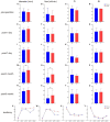
Results: A total of 52 patients with 54 operated hemispheres were enrolled, including 25 men and 27 women with an average age of 39 ± 14.3 years. Compared to preoperative values, the average blood flow of an STA graft at day 1 postoperation increased from 16.06 ± 12.47 to 117.47± 73.77 (mL/min), diameter increased from 1.14 ± 0.33 to 1.81 ± 0.30 (mm), PI dropped from 1.77 ± 0.42 to 0.76 ± 0.37, and RI dropped from 1.77 ± 0.42 to 0.50 ± 0.12. According to the Matsushima grade at 6 months after surgery, 30 hemispheres qualified as W group and 24 hemispheres as P group. Statistically significant differences were found between the two groups in diameter (p = 0.010) as well as flow (p = 0.017) at 3 months post-surgery. Flow also remained significantly different at 6 months after surgery (p = 0.014). Based on GEE logistic regression evaluation, the patients with higher levels of flow post-operation were more likely to have poorly-compensated collateral. ROC analysis showed that increased flow of ≥69.5 ml/min (p = 0.003; AUC = 0.74) or a 604% (p = 0.012; AUC = 0.70) increase at 3 months post-surgery compared with the pre-operative value is the cut-off point which had the highest Youden's index for predicting P group. Furthermore, a diameter at 3 months post-surgery that is ≥0.75 mm (p = 0.008; AUC = 0.71) or 52% (p =0.021; AUC = 0.68) wider than pre-operation also indicates a high risk of poor indirect collateral formation.
Conclusions: The hemodynamic of the STA graft changed significantly after combined bypass surgery. An increased flow of more than 69.5 ml/min at 3 months was a good predictive factor for poor neoangiogenesis in MMD patients treated with combined bypass surgery.
Keywords: Moyamoya; combined bypass surgery; forecast; hemodynamics; revascularization; ultrasonic.
Copyright © 2023 Chen, Wang, Wen, Wang, Long, Chen, Zhang, Li, Zhang, Pan, Feng, Qi and Wang.
Conflict of interest statement
The authors declare that the research was conducted in the absence of any commercial or financial relationships that could be construed as a potential conflict of interest.
Figures


References
-
- Sun J, Li Z-Y, Chen C, Ling C, Li H, Wang H. Postoperative neovascularization, cerebral hemodynamics, and clinical prognosis between combined and indirect bypass revascularization procedures in hemorrhagic Moyamoya disease. Clin Neurol Neurosurg. (2021) 208:106869. 10.1016/j.clineuro.2021.106869 - DOI - PubMed
-
- Zhang M, Raynald, Zhang D, Liu X, Wang R, Zhang Y, et al. Combined STA-MCA bypass and encephalodurosynangiosis versus encephalodurosynangiosis alone in adult hemorrhagic Moyamoya disease: a 5 -year outcome study. J Stroke Cerebrovasc Dis. (2020) 29:104811. 10.1016/j.jstrokecerebrovasdis.2020.104811 - DOI - PubMed
LinkOut - more resources
Full Text Sources
Research Materials
Miscellaneous

