. 2023 Feb 16:17:1053877.
doi: 10.3389/fnbeh.2023.1053877. eCollection 2023.

Enriched environment promotes post-stroke angiogenesis through astrocytic interleukin-17A

Affiliations

Enriched environment promotes post-stroke angiogenesis through astrocytic interleukin-17A

Xiuping Chen et al. Front Behav Neurosci. .

Abstract

Objective: Our previous studies have revealed that the protective effect of an enriched environment (EE) may be linked with astrocyte proliferation and angiogenesis. However, the relationship between astrocytes and angiogenesis under EE conditions still requires further study. The current research examined the neuroprotective effects of EE on angiogenesis in an astrocytic interleukin-17A (IL-17A)-dependent manner following cerebral ischemia/reperfusion (I/R) injury.

Methods: A rat model of ischemic stroke based on middle cerebral artery occlusion (MCAO) for 120 min followed by reperfusion was established, after which rats were housed in either EE or standard conditions. A set of behavior tests were conducted, including the modified neurological severity scores (mNSS) and the rotarod test. The infarct volume was evaluated by means of 2,3,5-Triphenyl tetrazolium chloride (TTC) staining. To evaluate the levels of angiogenesis, the protein levels of CD34 were examined by means of immunofluorescence and western blotting, while the protein and mRNA levels of IL-17A, vascular endothelial growth factor (VEGF), and the angiogenesis-associated factors interleukin-6 (IL-6), JAK2, and STAT3 were detected by western blotting and real-time quantitative PCR (RT-qPCR).

Results: We found that EE promoted functional recovery, reduced infarct volume, and enhanced angiogenesis compared to rats in standard conditions. IL-17A expression in astrocytes was also increased in EE rats. EE treatment increased the levels of microvascular density (MVD) and promoted the expression of CD34, VEGF, IL-6, JAK2, and STAT3 in the penumbra, while the intracerebroventricular injection of the IL-17A-neutralizing antibody in EE rats attenuated EE-mediated functional recovery and angiogenesis.

Conclusion: Our findings revealed a possible neuroprotective mechanism of astrocytic IL-17A in EE-mediated angiogenesis and functional recovery after I/R injury, which might provide the theoretical basis for EE in clinical practise for stroke patients and open up new ideas for the research on the neural repair mechanism mediated by IL-17A in the recovery phase of stroke.

Keywords: IL-17A; angiogenesis; astrocyte; cerebral ischemia/reperfusion; enriched environment.


Conflict of interest statement

The authors declare that the research was conducted in the absence of any commercial or financial relationships that could be construed as a potential conflict of interest.

Figures
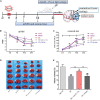
FIGURE 1
FIGURE 1
Schematic drawing of the experimental timeline and effect of enriched environment (EE) treatment on functional outcomes and infarct volume 21 days after middle cerebral artery occlusion (MCAO). (A) Timeline of behavioral testing and intracerebroventricular injection of interleukin-17A (IL-17A)-neutralizing mAb [2 μg, Intracerebroventricular (i.c.v.)] or IgG1 isotype after stroke. At 21 days after behavioral testing, the rats were sacrificed for tissue testing. (B) Evaluation of the neurological function of rats in the sham, MCAO, EE, EE + anti-IL-17A, and EE + isotype groups using modified neurological severity scores (mNSS) scores, n = 12 in each group. (C) The results of the rotarod test in five groups through measurement of time on the rod(s), n = 12 in each group. (D) Representative 2 mm coronal sections of each group. (E) Quantitative analysis of infarct volume percentage in each group. Sham-operated rats had no ischemic tissue (data not shown). Data are presented as mean ± SD. #P < 0.05, ##P < 0.01 vs. MCAO group; +P < 0.05, ++P < 0.01 vs. EE + anti-IL-17A group.
FIGURE 2
FIGURE 2
Enriched environment (EE) increased the protein and mRNA expression of interleukin-17A (IL-17A) in astrocytes at 21 days after middle cerebral artery occlusion (MCAO). (A) Double immunofluorescence imaging of IL-17A (red) cells co-expressing glial fibrillary acidic protein (GFAP) (green) in penumbra. Bar = 50 μm. (B) Quantitative analysis of double-labeled cells in penumbra, n = 6. (C) Quantitative analysis of IL-17A mRNA levels in penumbra, n = 3. (D,E) Western blotting and quantification illustrating increasing expression of IL-17A in the ischemic hemisphere, n = 3. Data are presented as mean ± SD. *P < 0.05, **P < 0.01 vs. sham group; #P < 0.05, ##P < 0.01 vs. MCAO group; ++P < 0.01 vs. EE + anti-IL-17A group.
FIGURE 3
FIGURE 3
Enriched environment (EE) promoted angiogenesis in penumbra in an interleukin-17A (IL-17A)-dependent manner in penumbra 21 days after middle cerebral artery occlusion (MCAO). (A) Representative images of CD34 immunostaining for each group. Bar = 50 μm. (B) Quantitative analysis of microvascular density (MVD) in the brain sections in the penumbra of each group, n = 3. (C,D) Western blotting and quantitative data for CD34 in the ischemic hemisphere, n = 3. Data are presented as mean ± SD. *P < 0.05, **P < 0.01 vs. sham group; ##P < 0.01 vs. MCAO group; ++P < 0.01 vs. EE + anti-IL-17A group.
FIGURE 4
FIGURE 4
Enriched environment (EE) treatment increased vascular endothelial growth factor (VEGF) protein and mRNA expression in penumbra in an interleukin-17A (IL-17A)-dependent manner 21 days after ischemic stroke. Western blotting (A) and quantification analysis (B) of VEGF expression in the five groups. (C) Quantitative analysis of VEGF mRNA levels in penumbra. Data (n = 3/group) are presented as mean ± SD. **P < 0.01 vs. sham group; ##P < 0.01 vs. MCAO group; ++P < 0.01 vs. EE + anti-IL-17A group.
FIGURE 5
FIGURE 5
Enriched environment (EE) treatment increased the protein and mRNA levels of the angiogenesis-associated factors IL-6, JAK2, and STAT3 in the penumbra in an interleukin-17A (IL-17A)-dependent manner 21 days after middle cerebral artery occlusion (MCAO). (A–D) Western blotting and quantification of angiogenesis-related proteins including IL-6, JAK2, and STAT3 in penumbra. (E–G) Quantitative analysis of IL-6, JAK2, and STAT3 mRNA levels in penumbra. Data (n = 3/group) are presented as mean ± SD. **P < 0.01 vs. sham group; ##P < 0.01 vs. MCAO group; ++P < 0.01 vs. EE + anti-IL-17A group.

References

    1. Abdel-Razik A., Mousa N., Elhelaly R., Elzehery R., Hasan A., Abdelsalam M., et al. (2020). Helicobacter pylori as an initiating factor of complications in patients with cirrhosis: A single-center observational study. Front. Med. (Lausanne) 7:96. 10.3389/fmed.2020.00096 - DOI - PMC - PubMed
    1. Aharoni R., Eilam R., Arnon R. (2021). Astrocytes in multiple sclerosis-essential constituents with diverse multifaceted functions. Int. J. Mol. Sci. 22:5904. - PMC - PubMed
    1. Chen J., Yu Y., Yuan Y., Zhang Y., Fan X., Yuan S., et al. (2017). Enriched housing promotes post-stroke functional recovery through astrocytic HMGB1-IL-6-mediated angiogenesis. Cell Death Discov. 3:17054. 10.1038/cddiscovery.2017.54 - DOI - PMC - PubMed
    1. Chen X., Zhang X., Liao W., Wan Q. (2017). Effect of physical and social components of enriched environment on astrocytes proliferation in rats after cerebral ischemia/reperfusion injury. Neurochem. Res. 42 1308–1316. - PubMed
    1. Chen X., Zhang X. (2022). Effect of prior exposure to enriched environment on cellular apoptosis after experimental stroke. Mol. Biol. Rep. 49 6541–6551. 10.1007/s11033-022-07494-y - DOI - PubMed