Review
. 2023 Feb 16:10:1106318.
doi: 10.3389/fmed.2023.1106318. eCollection 2023.

Lichen sclerosus: The 2023 update

Affiliations
Review

Lichen sclerosus: The 2023 update

David A De Luca et al. Front Med (Lausanne). .

Abstract

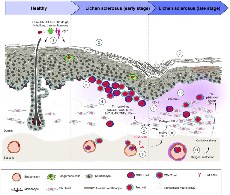
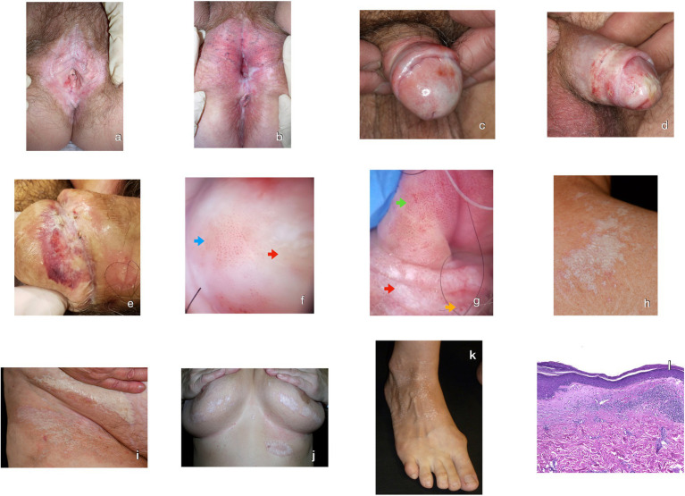
Lichen sclerosus (LS) is an underdiagnosed inflammatory mucocutaneous condition affecting the anogenital areas. Postmenopausal women are predominantly affected and, to a lesser extent, men, prepubertal children, and adolescents. The etiology of LS is still unknown. Hormonal status, frequent trauma and autoimmune diseases are well-known associations for LS, yet infections do not seem to be clear risk factors. LS pathogenesis involves factors such as a genetic predisposition and an immune-mediated Th1-specific IFNγ-induced phenotype. Furthermore, there is a distinct expression of tissue remodeling associated genes as well as microRNAs. Oxidative stress with lipid and DNA peroxidation provides an enabling microenvironment to autoimmunity and carcinogenesis. Circulating IgG autoantibodies against the extracellular matrix protein 1 and hemidesmosome may contribute to the progression of LS or simply represent an epiphenomenon. The typical clinical picture includes chronic whitish atrophic patches along with itching and soreness in the vulvar, perianal and penile regions. In addition to genital scarring, and sexual and urinary dysfunction, LS may also lead to squamous cell carcinoma. Disseminated extragenital LS and oral LS are also reported. The diagnosis is usually clinical; however, a skin biopsy should be performed in case of an unclear clinical picture, treatment failure or suspicion of a neoplasm. The gold-standard therapy is the long-term application of ultrapotent or potent topical corticosteroids and, alternatively, topical calcineurin inhibitors such as pimecrolimus or tacrolimus. Collectively, LS is a common dermatological disease with a so far incompletely understood pathogenesis and only limited treatment options. To foster translational research in LS, we provide here an update on its clinical features, pathogenesis, diagnosis and (emerging) treatment options.

Keywords: autoimmunity; balanitis xerotica obliterans; kraurosis vulvae; lichen sclerosus; white spot disease.


Conflict of interest statement

DT: Honoraria for participation on advisory boards, as a speaker and for consultancy from AbbVie, Almirall, Boehringer Ingelheim, Bristol Myers Squibb, Eli Lilly, Galapagos, Janssen, LEO Pharma, Morphosis, Novartis, Pfizer, Regeneron, Samsung, Sandoz, Sanofi Genzyme, and UCB Pharma; research grants received from LEO Pharma, and Novartis. The remaining authors declare that the research was conducted in the absence of any commercial or financial relationships that could be construed as a potential conflict of interest.

Figures

Figure 1
Figure 1
Schematic overview of lichen sclerosus pathogenesis. (1) Risk factors for LS; (2) The inflammatory early stage of LS is unspecific and shows a dermoepidermal interface band of mostly T-cells. (3) miR-155 is overexpressed in LS, stimulates the Th1 profile and the dermal sclerosis. (4) Th1 cytokines implicated in the pathogenesis of LS. (5) Lichenoid infiltrate of TCD4+, TCD8+, and Tregs cells in the upper dermis. (6) Abnormal expression of CD44 in the epidermis and lichenoid infiltrate. (7) The sclerotic late stage of LS presents an atrophic epidermis, with comedo-like plugs and a cleft in the stratum corneum, dermal sclerosis, reduction and dilation of dermal vessels and the appendages disappear. (8) The autoantibodies against EMC1 may activate MMP9, which subsequently activates TGF-β. (9) TGF-β and BMP2 induce the synthesis of collagen I and III in fibroblasts. (10) Galectin-7 induces the synthesis of collagen I and III. (11) Along with inflammation, the vessel sclerosis contributes to the oxidative stress in LS, leading to downregulation of tumor suppressors genes and overexpression of p53 in the skin, factors related with the development of skin carcinomas. The precise sequence of events and their interactions are only incompletely understood. References: BMP2: bone morphogenetic protein 2; EMC1: extracellular matrix protein 1; LS: lichen sclerosus; MMP9: metalloproteinase 9; TGF-β: tumor growth factor beta. TNF-α: Tumor necrosis factor alpha.
Figure 2
Figure 2
Clinical, dermoscopic and histological hallmarks of lichen sclerosus (LS). (A) Ivory-white wax-textured atrophic patches in the vulva. Agglutination, labial resorption, clitoral hood scarring. Fissures and erosions due to the narrowing of the vaginal introitus. (B) Erythema and anogenital scarring. Loss of the normal vulvar architecture with resorption of labia majora and minora. Ivory-white perineal spots perianal and scarring. (C) Hypopigmented macules, erythema and adhesions between glans and preputium. (D) Extensive scarring and atrophy of glans and partial loss of the balanopreputial sulcus. Ecchymotic and hypopigmented penile shaft. (E) Hemorrhagic variant of lichen sclerosus in glans penis over whitish scars, erosions and crusts. (F) Dermoscopy of LS in glans penis with structureless whitish and yellowish patches (red arrow), glomerular and dotted vessels (blue arrow). (G) Dermoscopy of LS in glans penis with penile intraepithelial neoplasia transformation. Glomerular vessels grouped over a pink and yellow surface, compatible with lichen sclerosus (green arrow). Structureless infiltrated whitish areas (red arrow) with glomerular and dotted vessels (yellow arrow), with histopathological confirmation of differentiated penile intraepithelial neoplasia. (H) Scapular ivory-white polygonal coalescing papules with adjacent erythema and comedo-like openings. (I) Whitish extensive macules, surrounded by hyperpigmentation, erythema and areas of ecchymosis on the groin. (J) Mammary and submammary ivory-white wax-textured atrophic coalescing spots with perilesional erythema and hyperpigmentation. (K) Small monomorph ivory-white wax-textured spots in extragenital lichen sclerosus. (L) The histopathology from a skin biopsy showing an atrophic epidermis with compact orthohyperkeratosis, hypergranulosis, degeneration of basal keratinocytes, and exocytosis. Under the thick basement membrane, there is a scarcely cellular zone of sclerosis. In the upper dermis, a superficial lichenoid inflammatory band and vasodilation with perivascular infiltrate are shown (H&E 40x).

References

    1. Hallopeau H. Leçons cliniques sur les maladies cutanées et syphilitiques. Union Med Can. (1887) 43:472.
    1. Kaufman RH, DiPaola GR, Friedrich EG, Jr, Hewitt J, Woodruff JD. New nomenclature for vulvar disease. J Cutan Pathol. (1976) 3:159–61. doi: 10.1111/j.1600-0560.1976.tb01105.x, PMID: - DOI - PubMed
    1. Papini M, Russo A, Simonetti O, Borghi A, Corazza M, Piaserico S, et al. . Mucous membrane disorders research group of SIDeMaST. Diagnosis and management of cutaneous and anogenital lichen sclerosus: recommendations from the Italian Society of Dermatology (SIDeMaST). Ital J Dermatol Venerol. (2021) 156:519–33. doi: 10.23736/S2784-8671.21.06764-X, PMID: - DOI - PubMed
    1. Schlosser BJ, Mirowski GW. Lichen sclerosus and lichen planus in women and girls. Clin Obstet Gynecol. (2015) 58:125–42. doi: 10.1097/GRF.0000000000000090 - DOI - PubMed
    1. Krapf JM, Mitchell L, Holton MA, Goldstein AT. Vulvar lichen Sclerosus: current perspectives. Int J Women's Health. (2020) 12:11–20. doi: 10.2147/IJWH.S191200 - DOI - PMC - PubMed