. 2023 Feb 17:14:1117821.
doi: 10.3389/fgene.2023.1117821. eCollection 2023.

Pathogenic gene variants in CCDC39, CCDC40, RSPH1, RSPH9, HYDIN, and SPEF2 cause defects of sperm flagella composition and male infertility

Affiliations

Pathogenic gene variants in CCDC39, CCDC40, RSPH1, RSPH9, HYDIN, and SPEF2 cause defects of sperm flagella composition and male infertility

I Aprea et al. Front Genet. .

Abstract

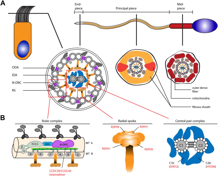
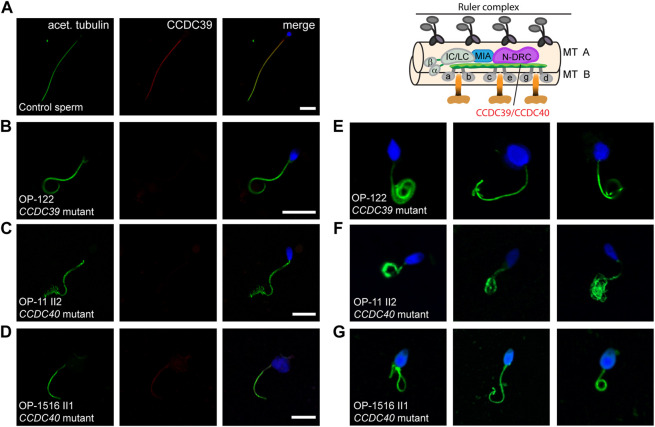
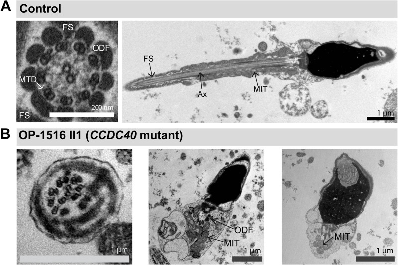
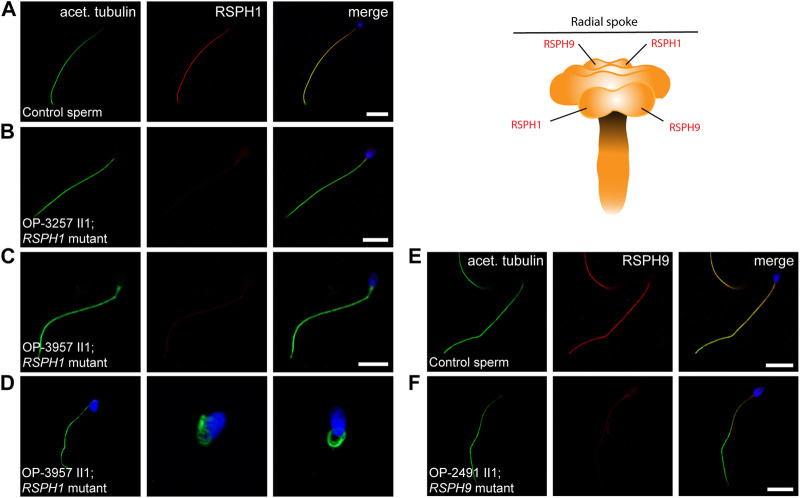
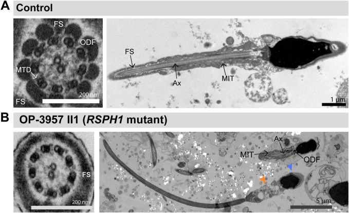
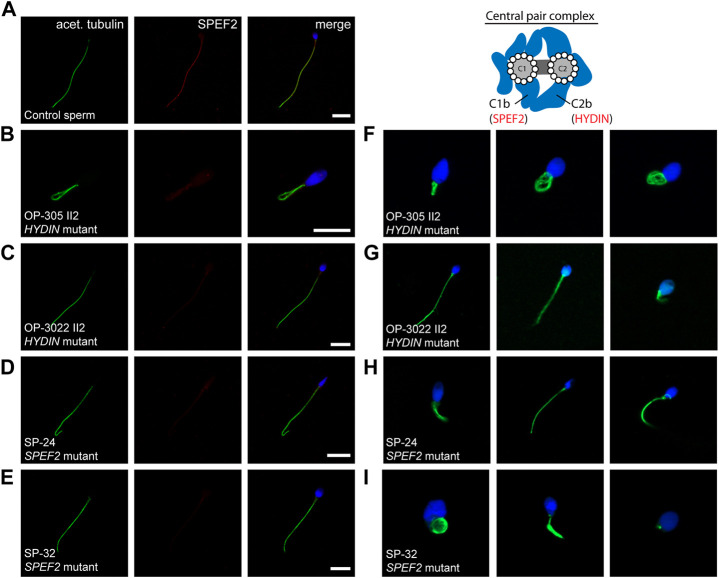
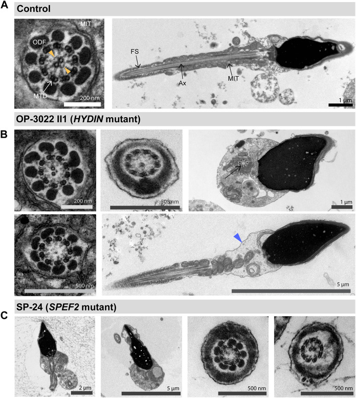
Primary Ciliary Dyskinesia (PCD) is a rare genetic disorder affecting the function of motile cilia in several organ systems. In PCD, male infertility is caused by defective sperm flagella composition or deficient motile cilia function in the efferent ducts of the male reproductive system. Different PCD-associated genes encoding axonemal components involved in the regulation of ciliary and flagellar beating are also reported to cause infertility due to multiple morphological abnormalities of the sperm flagella (MMAF). Here, we performed genetic testing by next generation sequencing techniques, PCD diagnostics including immunofluorescence-, transmission electron-, and high-speed video microscopy on sperm flagella and andrological work up including semen analyses. We identified ten infertile male individuals with pathogenic variants in CCDC39 (one) and CCDC40 (two) encoding ruler proteins, RSPH1 (two) and RSPH9 (one) encoding radial spoke head proteins, and HYDIN (two) and SPEF2 (two) encoding CP-associated proteins, respectively. We demonstrate for the first time that pathogenic variants in RSPH1 and RSPH9 cause male infertility due to sperm cell dysmotility and abnormal flagellar RSPH1 and RSPH9 composition. We also provide novel evidence for MMAF in HYDIN- and RSPH1-mutant individuals. We show absence or severe reduction of CCDC39 and SPEF2 in sperm flagella of CCDC39- and CCDC40-mutant individuals and HYDIN- and SPEF2-mutant individuals, respectively. Thereby, we reveal interactions between CCDC39 and CCDC40 as well as HYDIN and SPEF2 in sperm flagella. Our findings demonstrate that immunofluorescence microscopy in sperm cells is a valuable tool to identify flagellar defects related to the axonemal ruler, radial spoke head and the central pair apparatus, thus aiding the diagnosis of male infertility. This is of particular importance to classify the pathogenicity of genetic defects, especially in cases of missense variants of unknown significance, or to interpret HYDIN variants that are confounded by the presence of the almost identical pseudogene HYDIN2.

Keywords: CP-complex; MMAF; PCD; RS head; asthenozoospermia; axonemal ruler.


Conflict of interest statement

The authors declare that the research was conducted in the absence of any commercial or financial relationships that could be construed as a potential conflict of interest.

Figures

FIGURE 1
FIGURE 1
Schematic of the composition of motile 9 + 2 cilia and sperm flagella as well as representation of selected functional axonemal protein complexes involved in ciliary and flagellar beat regulation. (A) The upper panel shows a schematic representation of a ciliated respiratory epithelial cell (left) and a sperm cell (right). The sperm flagellum is divided into three regions; the mid piece, principal- and end piece. The middle panel shows cross sections through a respiratory cilia and the sperm flagellum. Both share a 9 + 2 axoneme composed of nine outer microtubule doublets (A- and B-tubule) surrounding a centered single pair of microtubules. Each outer A-tubule carries dynein based motor protein complexes, the outer- and inner dynein arms (respectively ODA and IDA). A nexin-link dynein regulatory complex (N-DRC) interconnects the outer doublets, while radial spoke (RS) complexes connect them to the central pair (CP) complex. In the sperm flagellar principal- and mid-piece the axoneme is additionally surrounded by accessory structures, such as the outer dense fibers (ODFs), the fibrous sheath (FS) and the mitochondrial (MT) sheath. (B) The lower panel shows schematics of the axonemal ruler, the RS and the CP complex. The ruler complex is representatively depicted in a longitudinal section of the axoneme. CCDC39 and CCDC40 form a heterodimer that is supposed to be located at the interface of the N-DRC and IDAs (composed of double headed IDA I1 with heavy chains α and β and the intermediate- and light chain (IC/LC) complex, and single headed IDAs a-e, and g) (Olbrich et al., 2015; Yamamoto et al., 2021). The RS complex is composed of a stalk and a head structure. RSPH1 and RSPH9, here highlighted, are known to be components of the RS head in human respiratory cilia (Frommer et al., 2015). The CP-complex is composed of two central microtubules connected by three intermicrotubular bridges. Several protein complexes are associated with the CP, e.g., the C1b and C2b projections, containing the proteins SPEF2 and HYDIN, respectively. In human respiratory cilia localization of SPEF2 depends on functional HYDIN (Cindrić et al., 2019).
FIGURE 2
FIGURE 2
CCDC40-mutant individuals display MMAF and CCDC39 is absent from CCDC39- and CCDC40-mutant sperm flagellar axonemes. Sperm cells from healthy control donors and infertile male individuals with pathogenic variants in CCDC39 and CCDC40 were co-stained with antibodies directed against acetylated α-tubulin (acet. tubulin; depicted in green) and CCDC39 (depicted in red). (A) In control sperm, immunoreactivity against CCDC39 (red) is observed along the entire flagella length. The CCDC39 signal co-localizes with the flagellar marker acet. tubulin (depicted in yellow in the merged image). (B–D) In sperm flagella of CCDC39-mutant individual OP-122 and CCDC40-mutant individuals OP-11 II2 and OP-1516 II1, no signal against CCDC39 is observed along the flagellar axoneme. (E–G) Sperm cells of CCDC39-mutant individual OP-122 and CCDC40-mutant individuals OP-11 II2 and OP-1516 II1 display morphological abnormalities, comprising short, bend and irregular flagella, as well as head defects. The schematic representatively shows the localization of the CCDC39/CCDC40 heterodimer. Nuclei were stained with Hoechst33342. Scale bars represent 10 µm.
FIGURE 3
FIGURE 3
TEM analyses of CCDC40-mutant sperm cells show morphological abnormalities and tubular disorganisation. (A) TEM micrographs of sperm samples from a healthy control donor, showing a flagellar cross section with a regular 9 + 2 axoneme of microtubule doublets (MTD), (left image), and a longitduinal section showing the axoneme (Ax) at the center, with regularly composed mitochondria (MIT) and fibrous sheath (FS), respectively in the flagellar mid piece and principal piece (right image). (B) CCDC40-mutant individual OP-1516 II1 shows morphological abnormalities of the sperm cells. Some sperm cells depict severe head malformations and lack proper flagella, displaying a cytoplasmic bag with unassembled peri-axonemal components, such as outer dense fibers (ODF) and mitochondria (right and middle image). In sperm flagella cross sections, axonemes show severe tubular disorganization (left image).
FIGURE 4
FIGURE 4
RSPH1 and RSPH9 mutations cause abnormal radial spoke head composition in sperm flagella. Sperm cells from healthy control donors and infertile male individuals with pathogenic variants in RSPH1 and RSPH9 were co-stained with antibodies directed against acetylated α-tubulin (acet. tubulin; depicted in green) and RSPH1 and RSPH9, respectively (both depicted in red). (A) In control sperm cells, immunoreactivity against RSPH1 (red) is observed along the entire flagellar length. The RSPH1 signal co-localizes with the flagellar marker acet. tubulin (depicted in yellow in the merged image). (B,C) In sperm flagella of RSPH1-mutant individuals OP-3257 II1 and OP-3957 II1, a severe reduction of the RSPH1 signal or no signal is observed along the flagellar axonemes. (D) Sperm cells of RSPH1-mutant individual OP-3957 II1 display morphological abnormalities, comprising bent flagella and sperm head defects consistent with MMAF. (E) In control sperm cells, RSPH9 (red) shows a panaxonemal staining pattern and co-localization with the flagellar marker acet. tubulin (depicted in yellow in the merged image). (F) In contrast, sperm flagella of the RSPH9-mutant individual OP-2491 II1 display a severe reduction of the RSPH9 signal. The schematic shows the putative localization of the RSPH1 and RSPH9 proteins in the radial spoke head complex. Nuclei were stained with Hoechst33342. Scale bars represent 10 µm.
FIGURE 5
FIGURE 5
TEM analyses of RSPH1-mutant sperm cells displayed morphological abnormalities of the midpiece and head region, but preservation of the 9 + 2 axonemal organization. (A) TEM micrographs of sperm samples from a healthy control donor, showing a flagellar cross section with a regular 9 + 2 axoneme of microtubule doublets (MTD), (left image), and a longitudinal section, with regularly arranged mitochondria (MIT) and fibrous sheath (FS), respectively in the flagellar mid piece and principal piece (right image). (B) TEM micrographs of sperm samples from the RSPH1-mutant individual OP-3957 II1 show morphological abnormalities with dysplasia of the flagellar midpiece and abnormal head shapes (respectively indicated by orange and blue arrowheads), or sperm with only a cytoplasmic bag containing unassembled axonemal (Ax) and peri-axonemal components, such as outer dense fibers (ODF) and MIT (right image). Some sperm flagellar cross sections also display a normal ultrastructure with a regularly disposed 9 + 2 axoneme (left image).
FIGURE 6
FIGURE 6
SPEF2 is absent or severely reduced in sperm flagellar axonemes of HYDIN- and SPEF2-mutant individuals, presenting with MMAF-associated infertility. Sperm cells from healthy control donors and infertile male individuals with pathogenic variants in HYDIN and SPEF2 were co-stained with antibodies directed against acetylated α-tubulin (acet. tubulin; depicted in green) and SPEF2 (depicted in red). (A) In control sperm cells, immunoreactivity against SPEF2 (red) is observed along the entire flagellar length. The SPEF2 signal co-localizes with the flagellar marker acet. tubulin (depicted in yellow in the merged image). (B–E) In sperm cells of both HYDIN-mutant individuals (OP-305 II2 and OP-3022 II2) and SPEF2-mutant individuals (SP-24 and SP-32), a severe reduction of the SPEF2 signal or no signal against SPEF2 was observed along the flagellar axoneme. (F–I) Sperm of both HYDIN-and SPEF2-mutant individuals display a MMAF phenotype with short, bent, coiled and irregular flagella. The schematic representatively shows the localization of HYDIN and SPEF2 within the central pair associated apparatus. Nuclei were stained with Hoechst33342. Scale bars represent 10 µm.
FIGURE 7
FIGURE 7
TEM analyses of HYDIN and SPEF2-deficient sperm displays morphological abnormalities. (A) TEM micrographs of sperm samples from a healthy control donor, showing a flagellar cross section with a regular 9 + 2 axoneme of microtubule doublets (MTD), (left image), and a longitudinal section showing the axoneme (Ax) at the center, with regularly arranged mitochondria (MIT) and fibrous sheath (FS), respectively in the flagellar mid piece and principal piece (right image). The orange arrowheads in the cross section indicate projections of the central pair associated apparatus. (B) HYDIN-mutant sperm display flagellar cross sections with either a normal 9 + 2 ultrastructure of the axoneme displaying less pronounced CP projections (upper left image), or 9 + 1 cross sections with only one central microtubule (upper middle image), or a 9 + 0 axoneme without the CP (lower left image). Morphological abnormalities of the sperm flagella are also observed, comprising sperm cells displaying a cytoplasmic bag with unassembled axonemal and peri-axonemal components (upper right image, arrow), or mid piece dysplasia (lower right image, indicate by a blue arrowhead). (C) SPEF2-mutant sperm cells also display short and irregular flagella (first left image), or a cytoplasmic bag with unassembled flagellar components (second left image). Flagellar cross sections display either a 9 + 0 structure without the CP (second from the right image), or a 9 + 2 structure (first from the right image).

Similar articles

Cited by

References

    1. Adzhubei I. A., Schmidt S., Peshkin L., Ramensky V. E., Gerasimova A., Bork P., et al. (2010). A method and server for predicting damaging missense mutations. Nat. Methods 7, 248–249. 10.1038/nmeth0410-248 - DOI - PMC - PubMed
    1. American Thoracic Society European Respiratory Society (2005). ATS/ERS recommendations for standardized procedures for the online and offline measurement of exhaled lower respiratory nitric oxide and nasal nitric oxide, 2005. Am. J. Respir. Crit. Care Med. 171, 912–930. 10.1164/rccm.200406-710ST - DOI - PubMed
    1. Antony D., Becker-Heck A., Zariwala M. A., Schmidts M., Onoufriadis A., Forouhan M., et al. (2013). Mutations in CCDC39 and CCDC40 are the major cause of primary ciliary dyskinesia with axonemal disorganization and absent inner dynein arms. Hum. Mutat. 34, 462–472. 10.1002/humu.22261 - DOI - PMC - PubMed
    1. Aprea I., Nöthe-Menchen T., Dougherty G. W., Raidt J., Loges N. T., Kaiser T., et al. (2021a). Motility of efferent duct cilia aids passage of sperm cells through the male reproductive system. Mol. Hum. Reprod. 27, gaab009. 10.1093/molehr/gaab009 - DOI - PMC - PubMed
    1. Aprea I., Raidt J., Höben I. M., Loges N. T., Nöthe-Menchen T., Pennekamp P., et al. (2021b). Defects in the cytoplasmic assembly of axonemal dynein arms cause morphological abnormalities and dysmotility in sperm cells leading to male infertility. PLoS Genet. 17, e1009306. 10.1371/journal.pgen.1009306 - DOI - PMC - PubMed