Review
. 2023 Feb 16:14:1150835.
doi: 10.3389/fphar.2023.1150835. eCollection 2023.

Natural products can modulate inflammation in intervertebral disc degeneration

Affiliations
Review

Natural products can modulate inflammation in intervertebral disc degeneration

Zongtai Liu et al. Front Pharmacol. .

Abstract

Intervertebral discs (IVDs) play a crucial role in maintaining normal vertebral anatomy as well as mobile function. Intervertebral disc degeneration (IDD) is a common clinical symptom and is an important cause of low back pain (LBP). IDD is initially considered to be associated with aging and abnormal mechanical loads. However, over recent years, researchers have discovered that IDD is caused by a variety of mechanisms, including persistent inflammation, functional cell loss, accelerated extracellular matrix decomposition, the imbalance of functional components, and genetic metabolic disorders. Of these, inflammation is thought to interact with other mechanisms and is closely associated with the production of pain. Considering the key role of inflammation in IDD, the modulation of inflammation provides us with new options for mitigating the progression of degeneration and may even cause reversal. Many natural substances possess anti-inflammatory functions. Due to the wide availability of such substances, it is important that we screen and identify natural agents that are capable of regulating IVD inflammation. In fact, many studies have demonstrated the potential clinical application of natural substances for the regulation of inflammation in IDD; some of these have been proven to have excellent biosafety. In this review, we summarize the mechanisms and interactions that are responsible for inflammation in IDD and review the application of natural products for the modulation of degenerative disc inflammation.

Keywords: inflammation; intervertebral disc; intervertebral disc degeneration; lower back pain; natural product.


Conflict of interest statement

The authors declare that the research was conducted in the absence of any commercial or financial relationships that could be construed as a potential conflict of interest.

Figures

FIGURE 1
FIGURE 1
A comparison of the gene expression of inflammatory factors between groups with different leukocyte levels (A). A comparison of gene expression of MMPs between groups with different leukocyte levels (B). A comparison of the concentrations of inflammatory factors between groups with different leukocyte levels (C). A comparison of the concentrations of MMPs between groups with different leukocyte levels (D). West-blot comparison of the production of NF-κB/p65 between groups with different leukocyte levels (E). “*” p < 0.05 compared to P-PRP or L-PRP with controls, “#” p < 0.05 compared to L-PRP with P-PRP. Reproduced with permission from a previous publication (Jia et al., 2018).
FIGURE 2
FIGURE 2
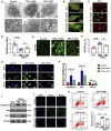
Comparison of human NP cells between groups under transmission electron microscopy (A) MitoTracker staining for mitochondria and phalloidin for cytoskeleton (B), scale bar = 10 μm. Comparison of JC-1 assay images between different groups (C), scale bar = 20 μm. Quantitative comparison of red and green fluorescence in JC-1 assays (D). Images of iNOS as determined by DCFDA staining (E), scale bar = 20 μm. Quantitative analysis of iNOS content (F). Comparison of NLRP3 staining in different groups (G), scale bar = 20 μm. Comparison of the caspase-3, Bax, and Bcl-2 mRNA levels in different groups (H). Western blot analysis of caspase-3, Bax and Bcl-2 in different groups (I). Images of TUNEL staining in different groups (J), scale bar = 100 μm. Comparison of the results of the number of apoptotic NPs, as measured by flow cytometry (K). “*” p < 0.05, “**” p < 0.01 and “***” p < 0.001. CST corticostatin. Reproduced with permission from a previous publication (Zhao et al., 2020).
FIGURE 3
FIGURE 3
Analysis of the enrichment of differential proteins in exosomes derived from normal CEP stem cells (N-exons) and exosomes derived from degenerate CEP stem cells (D-exons) using KEGG (A) Immunofluorescence images of LC3-B and cleaved caspase-3 in NP cells (B) Autophagosomes in each group were observed by transmission electron microscopy (C) Western blots and quantitative analysis of LC3B/A, Beclin-1, cleaved caspase-3, Bax, and Bcl-2 in each group. (D) “*” p < 0.05, “**” p < 0.01 and “***” p < 0.001. Reproduced with permission from a previous publication (Luo et al., 2021).
FIGURE 4
FIGURE 4
A comparison of the protein expression of MMP-3, TNF-α, and IL-1β in each group (A) A comparison of the protein expression of NF-κB in each group (B) Quantitative analysis of the protein levels of MMP-3 (C), TNF-α (D), IL-1β (E), and NF-κB (F), “*” p < 0.05. SB203580, a specific p38 MAPK inhibitor. Reproduced with permission from a previous publication (Ge et al., 2020).
FIGURE 5
FIGURE 5
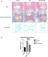
Images of IVD from rats in each treatment group stained with Safranin O/Fast Green and Alcian Blue in each group (A) A comparison of histological scores in each group (B), “****” p < 0.0001. Reproduced with permission from a previous publication (Wang et al., 2020b).
FIGURE 6
FIGURE 6
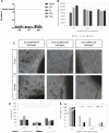
An evaluation of cytocompatibility of hydrogels containing different concentrations of curcumin by LDH (A) and Alamar blue (B). Optical microscope images of hydrogel cultured cells containing different concentrations of curcumin at 7 and 14 days (C). The levels of IL-8 (D) and TNF-α (E) produced by cells when exposed to hydrogels with different concentrations of curcumin. Reproduced with permission from a previous publication (Zamboni et al., 2022).

Similar articles

Cited by

References

    1. Afonina I. S., Zhong Z., Karin M., Beyaert R. (2017). Limiting inflammation-the negative regulation of NF-κB and the NLRP3 inflammasome. Nat. Immunol. 18, 861–869. 10.1038/ni.3772 - DOI - PubMed
    1. Al-Warhi T., Sabt A., Elkaeed E. B., Eldehna W. M. (2020). Recent advancements of coumarin-based anticancer agents: An up-to-date review. Bioorg Chem. 103, 104163. 10.1016/j.bioorg.2020.104163 - DOI - PubMed
    1. Arenas-Jal M., Suñé-Negre J. M., GarcíA-Montoya E. (2020). Coenzyme Q10 supplementation: Efficacy, safety, and formulation challenges. Compr. Rev. Food Sci. Food Saf. 19, 574–594. 10.1111/1541-4337.12539 - DOI - PubMed
    1. Ashinsky B. G., Bonnevie E. D., Mandalapu S. A., Pickup S., Wang C., Han L., et al. (2020). Intervertebral disc degeneration is associated with aberrant endplate remodeling and reduced small molecule transport. J. Bone Min. Res. 35, 1572–1581. 10.1002/jbmr.4009 - DOI - PMC - PubMed
    1. Atay O., Skotheim J. M. (2017). Spatial and temporal signal processing and decision making by MAPK pathways. J. Cell Biol. 216, 317–330. 10.1083/jcb.201609124 - DOI - PMC - PubMed