

Myocardial Infarction Due to Paradoxical Thromboembolism Originating From Distal Lower Extremity Deep Vein Thrombosis (LEDVT)
- PMID: 36874327
- PMCID: PMC9981484
- DOI: 10.7759/cureus.34592
Myocardial Infarction Due to Paradoxical Thromboembolism Originating From Distal Lower Extremity Deep Vein Thrombosis (LEDVT)
Abstract
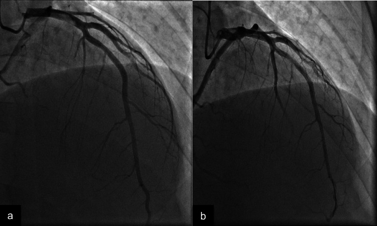
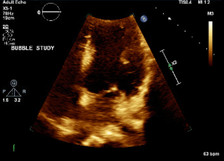
Paradoxical embolism (PDE) originates in the venous system and ends up in the arterial circulation via cardiac or pulmonary shunts. Cases of PDE from venous thrombosis resulting in acute myocardial infarctions (MIs) are seldom reported in the literature. Diagnoses can often be missed if further workups are not pursued in patients without any underlying risk factors for coronary artery disease (CAD). Here, we report a case of a paradoxical embolus that crossed the patent foramen ovale (PFO), causing ST-elevation MI (STEMI) from an embolized venous thrombus originating in the left distal posterior tibial vein.
Keywords: echocardiogram; myocardial infarction; paradoxical emboli; patent foramen ovale; thrombus.
Copyright © 2023, Josephs et al.
Conflict of interest statement
The authors have declared that no competing interests exist.
Figures

References
-
- Paradoxical embolism causing myocardial infarction. Nguyen R, Morales-Mangual C, Gu S, et al. J Med Cases. 2017;8:365–367.
-
- Hakman EN, Cowling KM. Treasure Island, FL: StatPearls; 2022. Paradoxical Embolism. - PubMed
Publication types
LinkOut - more resources
Full Text Sources
Miscellaneous