

Selfrec-Net: self-supervised deep learning approach for the reconstruction of Cherenkov-excited luminescence scanned tomography
- PMID: 36874507
- PMCID: PMC9979688
- DOI: 10.1364/BOE.480429
Selfrec-Net: self-supervised deep learning approach for the reconstruction of Cherenkov-excited luminescence scanned tomography
Abstract
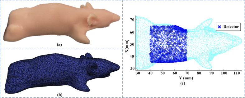
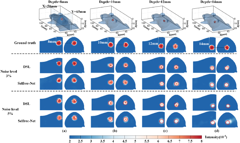
As an emerging imaging technique, Cherenkov-excited luminescence scanned tomography (CELST) can recover a high-resolution 3D distribution of quantum emission fields within tissue using X-ray excitation for deep penetrance. However, its reconstruction is an ill-posed and under-conditioned inverse problem because of the diffuse optical emission signal. Deep learning based image reconstruction has shown very good potential for solving these types of problems, however they suffer from a lack of ground-truth image data to confirm when used with experimental data. To overcome this, a self-supervised network cascaded by a 3D reconstruction network and the forward model, termed Selfrec-Net, was proposed to perform CELST reconstruction. Under this framework, the boundary measurements are input to the network to reconstruct the distribution of the quantum field and the predicted measurements are subsequently obtained by feeding the reconstructed result to the forward model. The network was trained by minimizing the loss between the input measurements and the predicted measurements rather than the reconstructed distributions and the corresponding ground truths. Comparative experiments were carried out on both numerical simulations and physical phantoms. For singular luminescent targets, the results demonstrate the effectiveness and robustness of the proposed network, and comparable performance can be attained to a state-of-the-art deep supervised learning algorithm, where the accuracy of the emission yield and localization of the objects was far superior to iterative reconstruction methods. Reconstruction of multiple objects is still reasonable with high localization accuracy, although with limits to the emission yield accuracy as the distribution becomes more complex. Overall though the reconstruction of Selfrec-Net provides a self-supervised way to recover the location and emission yield of molecular distributions in murine model tissues.
© 2023 Optica Publishing Group under the terms of the Optica Open Access Publishing Agreement.
Conflict of interest statement
The authors have no relevant financial interests in this work.
Figures


Similar articles
-
X-ray Cherenkov-luminescence tomography reconstruction with a three-component deep learning algorithm: Swin transformer, convolutional neural network, and locality module.J Biomed Opt. 2023 Feb;28(2):026004. doi: 10.1117/1.JBO.28.2.026004. Epub 2023 Feb 16. J Biomed Opt. 2023. PMID: 36818584 Free PMC article.
-
Cherenkov-excited luminescence scanned imaging using scanned beam differencing and iterative deconvolution in dynamic plan radiation delivery in a human breast phantom geometry.Med Phys. 2019 Jul;46(7):3067-3077. doi: 10.1002/mp.13545. Epub 2019 May 18. Med Phys. 2019. PMID: 30980725 Free PMC article.
-
Robust reconstruction of fluorescence molecular tomography based on adaptive adversarial learning strategy.Phys Med Biol. 2023 Feb 10;68(4). doi: 10.1088/1361-6560/acb638. Phys Med Biol. 2023. PMID: 36696695
-
An encoder-decoder network for direct image reconstruction on sinograms of a long axial field of view PET.Eur J Nucl Med Mol Imaging. 2022 Nov;49(13):4464-4477. doi: 10.1007/s00259-022-05861-2. Epub 2022 Jul 11. Eur J Nucl Med Mol Imaging. 2022. PMID: 35819497
-
Cone beam x-ray luminescence computed tomography: a feasibility study.Med Phys. 2013 Mar;40(3):031111. doi: 10.1118/1.4790694. Med Phys. 2013. PMID: 23464291
References
-
- Pogue B. W., Feng J., LaRochelle E. P., Brůža P., Lin H., Zhang R., Shell J. R., Dehghani H., Davis S. C., Vinogradov S. A., Gladstone D. J., Jarvis L. A., “Map of in vivo oxygen pressure with submillimeter resolution and nanomolar sensitivity enabled buy Cherenkov-exited luminescence scanned imaging,” Nat. Biomed. Eng. 2(4), 254–264 (2018).10.1038/s41551-018-0220-3 - DOI - PMC - PubMed
LinkOut - more resources
Full Text Sources
Research Materials