Review
. 2023 Feb 16:14:1131588.
doi: 10.3389/fimmu.2023.1131588. eCollection 2023.

Ongoing involvers and promising therapeutic targets of hepatic fibrosis: The hepatic immune microenvironment

Affiliations
Review

Ongoing involvers and promising therapeutic targets of hepatic fibrosis: The hepatic immune microenvironment

Nana Zhang et al. Front Immunol. .

Abstract

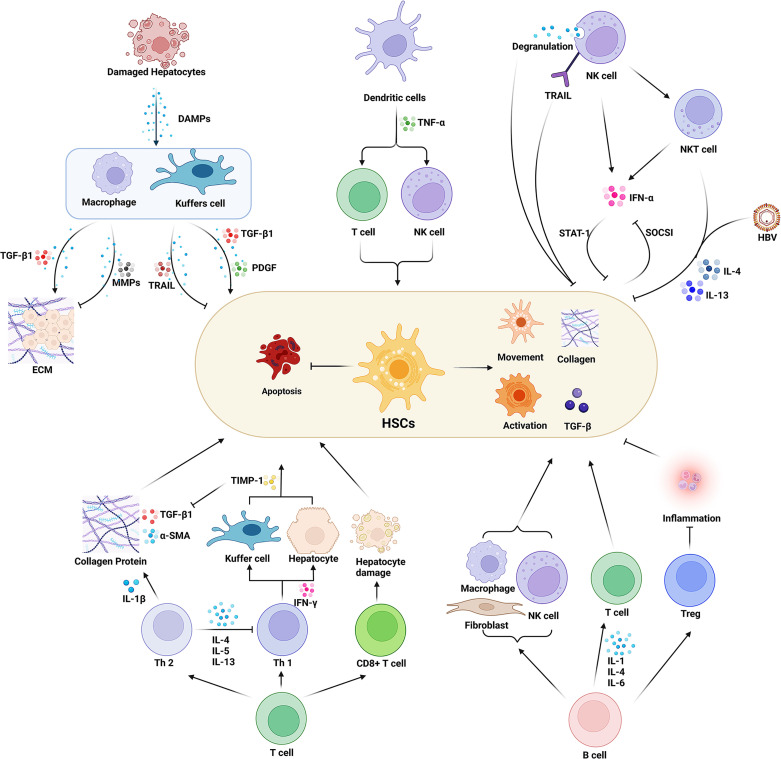
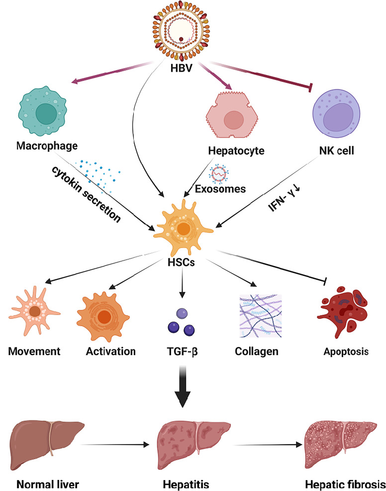
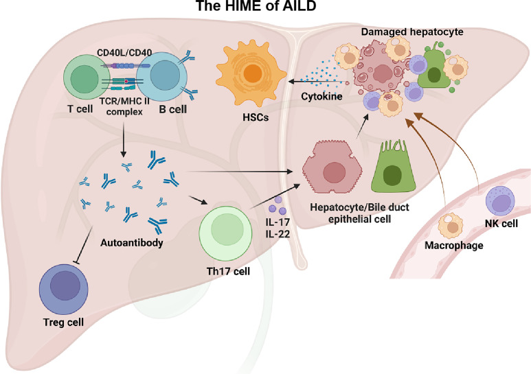
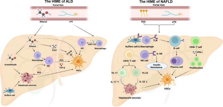
Hepatic fibrosis is often secondary to chronic inflammatory liver injury. During the development of hepatic fibrosis, the damaged hepatocytes and activated hepatic stellate cells (HSCs) caused by the pathogenic injury could secrete a variety of cytokines and chemokines, which will chemotactic innate and adaptive immune cells of liver tissue and peripheral circulation infiltrating into the injury site, mediating the immune response against injury and promoting tissue reparation. However, the continuous release of persistent injurious stimulus-induced inflammatory cytokines will promote HSCs-mediated fibrous tissue hyperproliferation and excessive repair, which will cause hepatic fibrosis development and progression to cirrhosis even liver cancer. And the activated HSCs can secrete various cytokines and chemokines, which directly interact with immune cells and actively participate in liver disease progression. Therefore, analyzing the changes in local immune homeostasis caused by immune response under different pathological states will greatly enrich our understanding of liver diseases' reversal, chronicity, progression, and even deterioration of liver cancer. In this review, we summarized the critical components of the hepatic immune microenvironment (HIME), different sub-type immune cells, and their released cytokines, according to their effect on the development of progression of hepatic fibrosis. And we also reviewed and analyzed the specific changes and the related mechanisms of the immune microenvironment in different chronic liver diseases.Moreover, we retrospectively analyzed whether the progression of hepatic fibrosis could be alleviated by modulating the HIME.We aimed to elucidate the pathogenesis of hepatic fibrosis and provide the possibility for exploring the therapeutic targets for hepatic fibrosis.

Keywords: chronic liver diseases; hepatic fibrosis; hepatic immune microenvironment; hepatic stellate cells (HSCs); immune cells.


Conflict of interest statement

The authors declare that the research was conducted in the absence of any commercial or financial relationships that could be construed as a potential conflict of interest.

Figures

Figure 1
Figure 1
Effect of various immune cells in the hepatic immune microenvironment (HIME) on hepatic fibrosis. Various immune cells affect the process of hepatic fibrosis by promoting or inhibiting the function of HSCs. Macrophages play a dual role in this process. The peripheral macrophages and the resident liver macrophages (Kuffer cells) are activated under the damage-associated molecular pattern released from the damaged hepatocytes during injury or inflammatory conditions. Activated macrophages secrete proinflammatory factors such as TGF-β to promote ECM formation and activate HSCs. On the other hand, macrophages can also inhibit hepatic fibrosis by expressing MMPs and TRAIL. Dendritic cells secrete TNF-α to activate HSCs by enhancing the immune function of T cells and NK cells. And the activated NK cells inhibit the function of HSCs and prevent hepatic fibrosis progression. While NK cells also can directly damage HSCs through degranulation and up-regulation of TRAIL expression. Moreover, it can secrete INF-γ, and synergize NKT cells to inhibit HSCs activation through the STAT signaling pathway. However, HSCs can inhibit INF-γ secretion of NK cells via the SOCSI pathway. The balance of Th1/Th2 cells plays an important role in the process of hepatic fibrosis. Th1 cells secrete INF-γ to increase the expression of TIMP in macrophages and inhibit the activity of HSCs. Th2 cells can secrete a variety of cytokines to activate HSCs and inhibit the HSCs’ apoptosis as well as the activity of Th1 cells. CD8+ T cells secrete cytotoxic factors to mediate hepatocyte injury, thereby activating HSCs. B cells regulate macrophages, fibroblasts, and T cells and promote HSCs activation by secreting interleukins. And B cells are also able to regulate Treg cells to inhibit inflammatory responses and HSCs’ activation. Created with BioRender.com.
Figure 2
Figure 2
The HIME of HBV-induced viral hepatitis. The hepatitis virus replicates within hepatocytes and is secreted into the extracellular space via e exosomes. Viral antigens can exert multiple effects in HSCs to promote hepatic fibrosis. In addition, the virus can be recognized by macrophages and NK cells, which will up-regulate or down-regulate the secretion of cytokines to activate HSCs. Subsequently, HSCs activation aggravates hepatitis and promotes the progression of hepatic fibrosis. Created with BioRender.com.
Figure 3
Figure 3
The HIME of AILD. Autoimmune liver disease happens when a large number of autoantigens are present in the liver. With the assistance of T cells, B cells can generate a large number of autoantibodies that recognize these autoantigens. These autoantibodies can impair the function of Treg cells and lead to autoimmune damage. In addition, autoantibodies can directly, or indirectly through Th17 cells, exert an effect on targeted cells (e.g., hepatocytes, biliary epithelial cells) and recruit peripheral macrophages and NK cells, causing liver injury. These injured cells release large amounts of cytokines thereby driving the HSCs activation. Created with BioRender.com.
Figure 4
Figure 4
The HIME of ALD and NAFLD. In ALD, ethanol is absorbed through the intestine and entry the portal vein to the liver. Ethanol can stimulate M2 macrophage polarization that secretes reactive oxygen species and TGF-β to promote hepatic fibrosis. Ethanol also increases P2X7R(P2X7 receptor) expression on HSCs, and P2X7 can promote HSCs activation. Acetaldehyde, an intermediate product of ethanol, induces hepatocyte necrosis. And necrotic hepatocytes directly activate HSCs and release ROS to activate M1 macrophages. Additionally, ethanol can disrupt the intestinal barrier, causing LPS to enter the liver via the portal vein and induce M2 macrophage polarization. In NAFLD, free fatty acid and LPS from the peripheral circulation induce TLR expression in macrophages, transform B cells into plasma cells, and secrete antibodies, which activate HSCs activation. In addition, free fatty acid and LPS are also able to stimulate CD4+ T cells, regulate the balance of Th17/Th22 cells, and induce hepatocyte necrosis by increasing IL-17 and reducing IL-22. Moreover, CD8+ T cells and NK cells can also promote HSCs activation under this HIME. Created with BioRender.com.

Similar articles

Cited by

References

    1. Hernandez-Gea V, Friedman SL. Pathogenesis of liver fibrosis. Annu Rev Pathol (2011) 6:425–56. doi: 10.1146/annurev-pathol-011110-130246 - DOI - PubMed
    1. Dhar D, Baglieri J, Kisseleva T, Brenner DA. Mechanisms of liver fibrosis and its role in liver cancer. Exp Biol Med (Maywood N.J.) (2020) 245:96–108. doi: 10.1177/1535370219898141 - DOI - PMC - PubMed
    1. Aydın MM, Akçalı KC. Liver fibrosis. Turkish J Gastroenterol (2018) 29:14–21. doi: 10.5152/tjg.2018.17330 - DOI - PMC - PubMed
    1. Deng Y, Xia B, Chen Z, Wang F, Lv Y, Chen G. Stem cell-based therapy strategy for hepatic fibrosis by targeting intrahepatic cells. Stem Cell Rev Rep (2021) 18:77–93. doi: 10.1007/s12015-021-10286-9 - DOI - PubMed
    1. Tsuchida T, Friedman SL. Mechanisms of hepatic stellate cell activation. Nat Rev Gastroenterol Hepatol (2017) 14:397–411. doi: 10.1038/nrgastro.2017.38 - DOI - PubMed

Publication types