Review
. 2023 Feb 15:15:1047897.
doi: 10.3389/fnagi.2023.1047897. eCollection 2023.

Potential roles of the endoplasmic reticulum stress pathway in amyotrophic lateral sclerosis

Affiliations
Review

Potential roles of the endoplasmic reticulum stress pathway in amyotrophic lateral sclerosis

Yu-Mi Jeon et al. Front Aging Neurosci. .

Abstract

The endoplasmic reticulum (ER) is a major organelle involved in protein quality control and cellular homeostasis. ER stress results from structural and functional dysfunction of the organelle, along with the accumulation of misfolded proteins and changes in calcium homeostasis, it leads to ER stress response pathway such as unfolded protein response (UPR). Neurons are particularly sensitive to the accumulation of misfolded proteins. Thus, the ER stress is involved in neurodegenerative diseases such as Alzheimer's disease, Parkinson's disease, prion disease and motor neuron disease (MND). Recently, the complex involvement of ER stress pathways has been demonstrated in experimental models of amyotrophic lateral sclerosis (ALS)/MND using pharmacological and genetic manipulation of the unfolded protein response (UPR), an adaptive response to ER stress. Here, we aim to provide recent evidence demonstrating that the ER stress pathway is an essential pathological mechanism of ALS. In addition, we also provide therapeutic strategies that can help treat diseases by targeting the ER stress pathway.

Keywords: amyotrophic lateral sclerosis; endoplasmic reticulum stress; motor neuron disease; therapeutic target; unfolded protein response.


Conflict of interest statement

The authors declare that the research was conducted in the absence of any commercial or financial relationships that could be construed as a potential conflict of interest.

Figures

Figure 1
Figure 1
Unfolded protein response (UPR) pathway. In normal condition, GRP78/BiP interact with UPR sensors (PERK, ATF6, and IRE1). When misfolded or unfolded proteins accumulated in ER, GRP78/BiP dissociated from UPR sensors and can bind to misfolded or unfolded proteins. (1) Protein kinase RNA-like endoplasmic reticulum kinase (PERK) autophosphorylates and induces eIF2α phosphorylation. (2) Activating transcription Factor 6 (ATF6) moves into Golgi apparatus and cleaved by S1P and S2P. Cleaved ATF6 moves into nucleus, activates UPR genes. (3) IRE1 autophosphorylates and splices XBP1.
Figure 2
Figure 2
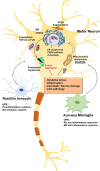
Relationship between endoplasmic reticulum (ER) stress and amyotrophic lateral sclerosis (ALS)-associated pathology. Misfolded proteins accumulate in the ER lumen and this accumulation contributes to ER stress. ER stress triggers the UPR to restore protein homeostasis. However, chronic stress leads to a cascade of intracellular death and inflammatory signal. Some nuclear ALS disease proteins such as TAR DNA-binding protein 43 (TDP-43) or fused in sarcoma (FUS) are depleted in the nucleus and accumulate in the cytoplasm in pathologic condition. Pathologic TDP-43 or FUS are existed as hyperphosphorylated and ubiquitinated form. Persistent activation of the stress response leads to ER dysfunction and ubiquitin proteasome system (UPS) impairment, leading to reactive oxygen species (ROS) and RNS release in mitochondria. This eventually leads to insoluble cytoplasmic aggregation and impairment of mitochondrial and ER function. Furthermore, UPR-related genes are upregulated in astrocytes and microglia along with inflammatory genes. Specifically, UPR activation reduces the ability of glia to support synapse and is associated with neuronal cell death. In addition, glia could transmit ER stress to neurons, which in turn may exacerbate ER stress mediated neuronal damage.

References

    1. Al-Saif A., Al-Mohanna F., Bohlega S. (2011). A mutation in sigma-1 receptor causes juvenile amyotrophic lateral sclerosis. Ann. Neurol. 70, 913–919. doi: 10.1002/ana.22534, PMID: - DOI - PubMed
    1. Ash P. E., Zhang Y. J., Roberts C. M., Saldi T., Hutter H., Buratti E., et al. . (2010). Neurotoxic effects of TDP-43 overexpression in C. elegans. Hum. Mol. Genet. 19, 3206–3218. doi: 10.1093/hmg/ddq230, PMID: - DOI - PMC - PubMed
    1. Ashford B. A., Boche D., Cooper-Knock J., Heath P. R., Simpson J. E., Highley J. R. (2021). Review: microglia in motor neuron disease. Neuropathol. Appl. Neurobiol. 47, 179–197. doi: 10.1111/nan.12640, PMID: - DOI - PubMed
    1. Atkin J. D., Farg M. A., Turner B. J., Tomas D., Lysaght J. A., Nunan J., et al. . (2017). Withdrawal: induction of the unfolded protein response in familial amyotrophic lateral sclerosis and association of protein-disulfide isomerase with superoxide dismutase 1. J. Biol. Chem. 292:12007. doi: 10.1074/jbc.A117.603393, PMID: - DOI - PMC - PubMed
    1. Bellezza I., Grottelli S., Mierla A. L., Cacciatore I., Fornasari E., Roscini L., et al. . (2014). Neuroinflammation and endoplasmic reticulum stress are coregulated by cyclo(his-pro) to prevent LPS neurotoxicity. Int. J. Biochem. Cell Biol. 51, 159–169. doi: 10.1016/j.biocel.2014.03.023, PMID: - DOI - PubMed