. 2022 May 9;2(5):330-341.
doi: 10.1158/2767-9764.CRC-22-0083. eCollection 2022 May.

Redirecting T-cell Activity with Anti-BCMA/Anti-CD3 Bispecific Antibodies in Chronic Lymphocytic Leukemia and Other B-cell Lymphomas

Affiliations

Redirecting T-cell Activity with Anti-BCMA/Anti-CD3 Bispecific Antibodies in Chronic Lymphocytic Leukemia and Other B-cell Lymphomas

Anne W J Martens et al. Cancer Res Commun. .

Abstract

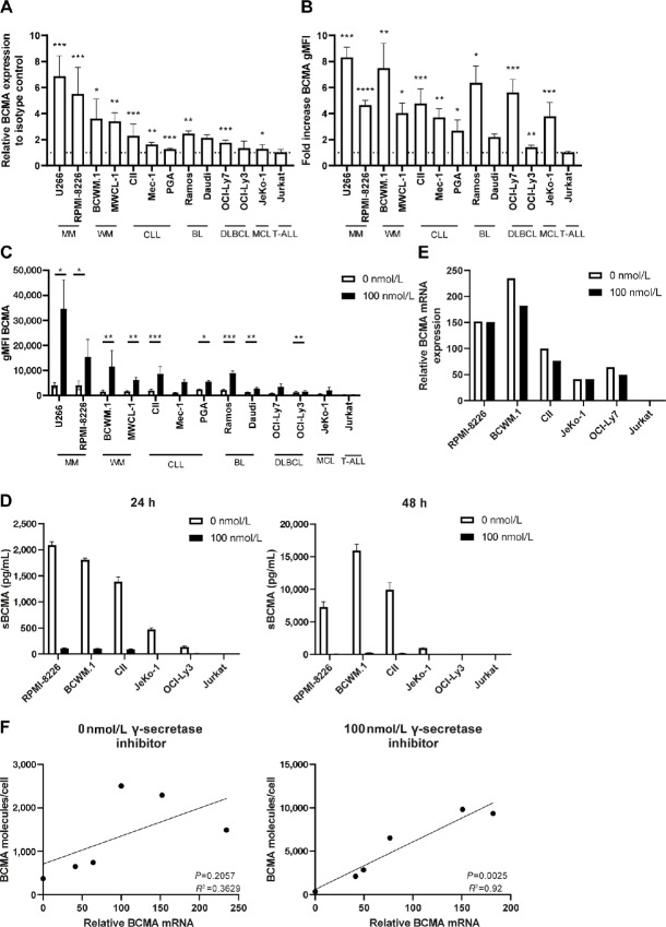
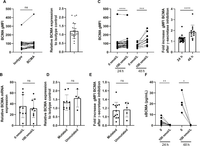
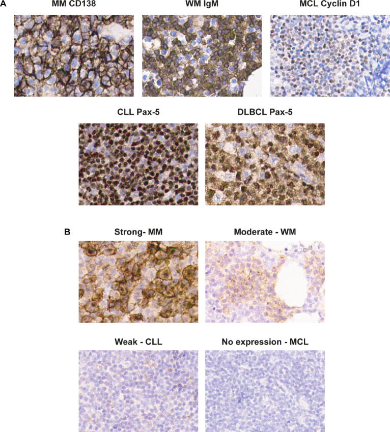
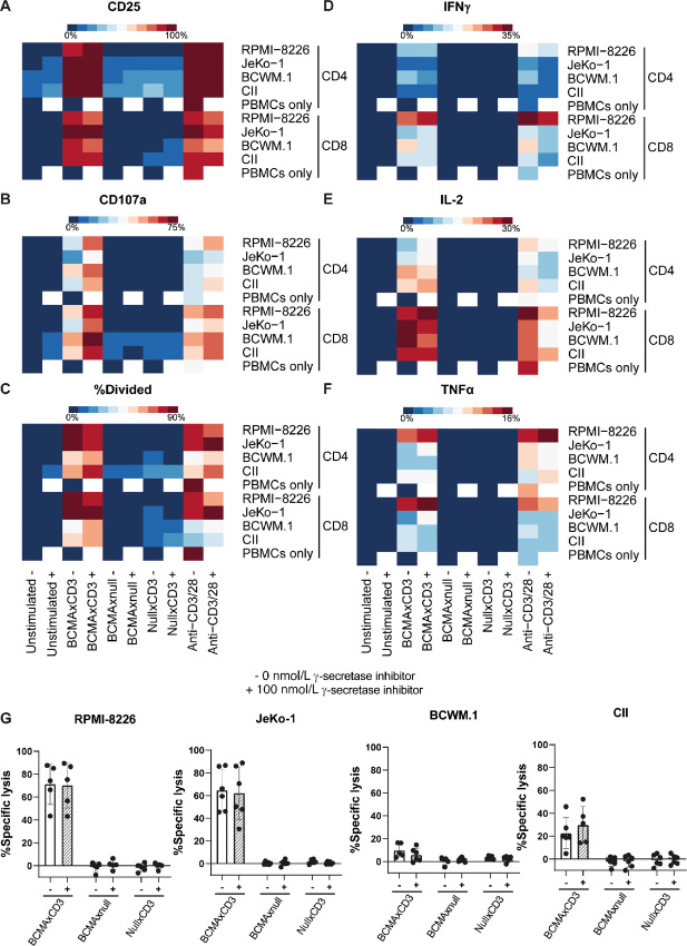
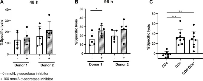
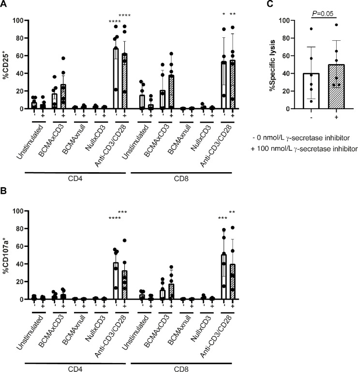
T-cell redirecting bispecific antibodies hold high promise for treatment of B-cell malignancies. B-cell maturation antigen (BCMA) exhibits high expression on normal and malignant mature B cells including plasma cells, which can be enhanced by inhibition of γ-secretase. BCMA is considered a validated target in multiple myeloma but whether mature B-cell lymphomas can be targeted by the BCMAxCD3 T-cell redirector teclistamab is currently unknown. BCMA expression on B-cell non-Hodgkin lymphoma and primary chronic lymphocytic leukemia (CLL) cells was assessed by flow cytometry and/or IHC. To assess teclistamab efficacy, cells were treated with teclistamab in presence of effector cells with/without γ-secretase inhibition. BCMA could be detected on all tested mature B-cell malignancy cell lines, while expression levels varied per tumor type. γ-secretase inhibition universally increased BCMA surface expression. These data were corroborated in primary samples from patients with Waldenstrom's macroglobulinemia, CLL, and diffuse large B-cell lymphoma. Functional studies with the B-cell lymphoma cell lines revealed teclistamab-mediated T-cell activation, proliferation, and cytotoxicity. This was independent of the level of BCMA expression, but generally lower in mature B-cell malignancies compared with multiple myeloma. Despite low BCMA levels, healthy donor T cells and CLL-derived T cells induced lysis of (autologous) CLL cells upon addition of teclistamab. These data show that BCMA is expressed on various B-cell malignancies and that lymphoma cell lines and primary CLL can be targeted using teclistamab. Further studies to understand the determinants of response to teclistamab are required to identify which other diseases might be suitable for teclistamab targeting.

Significance: Besides reported BCMA expression on multiple myeloma, we demonstrate BCMA can be detected and enhanced using γ-secretase inhibition on cell lines and primary material of various B-cell malignancies. Furthermore, using CLL we demonstrate that low BCMA-expressing tumors can be targeted efficiently using the BCMAxCD3 DuoBody teclistamab.


Conflict of interest statement

N.W.C.J. van de Donk reports grants and other from Janssen Pharmaceuticals during the conduct of the study; grants and other from Amgen, Celgene/BMS, Novartis, Cellectis; other from Takeda, Roche, Bayer, Adaptive, and Servier outside the submitted work. H.C. Adams reports other from Janssen R&D outside the submitted work; in addition, H.C. Adams has a patent to PRD4087 issued. E. Eldering reports other from Janssen during the conduct of the study; grants from Janssen outside the submitted work. R. Verona reports other from Janssen R&D during the conduct of the study; other from Janssen R&D outside the submitted work; in addition, R. Verona has a patent to 63/194,470 pending. A.P. Kater reports grants from Janssen during the conduct of the study; grants and other from Abbvie, Genentech, AstraZeneca, BMS, and other from LAVA outside the submitted work; in addition, A.P. Kater has a patent to BCMA-bispecific for lymphoma pending. No other disclosures were reported.

Figures

FIGURE 1
FIGURE 1
BCMA is expressed by different B-cell malignancies and can be enhanced by γ-secretase inhibition. A, B-cell malignancy cell lines were cultured and basal levels of BCMA were assessed by flow cytometry and compared with isotype controls (n = 3–8). Dotted line indicates no increase compared with isotype control. B, Cell lines were treated for 24–48 hours with 100 nmol/L γ-secretase inhibitor or with medium control and BCMA was assessed by flow cytometry. Values are represented as fold increase compared with 0 nmol/L γ-secretase inhibitor (n = 3–12). Dotted line indicates no increase compared with 0 nmol/L γ-secretase inhibitor. C, Cell lines were cultured for 24 hours with 100 nmol/L γ-secretase inhibitor or with medium control and BCMA was assessed by flow cytometry. Values are molecules/cell as quantified using PE Fluorescence Quantitation Kit (n = 3–12). D, Assessment of soluble BCMA by ELISA in supernatants of B-cell malignancy cell lines after treatment with 0 or 100 nmol/L γ-secretase inhibitor for 24–48 hours (n = 2). E, Assessment of BCMA mRNA relative to GAPDH control by qPCR after B-cell malignancy cell lines were treated without or with 100 nmol/L γ-secretase inhibitor for 24 hours. F, Correlation between BCMA membrane expression and BCMA mRNA expression in B-cell malignancy cell lines without γ-secretase inhibition or with 100 nmol/L γ-secretase inhibition for 24 hours. The P value was calculated by paired t test or Wilcoxon test (A–C) or simple linear regression (E). In A, P values were calculated compared with isotype controls. In B, P values were calculated compared with 0 nmol/L γ-secretase inhibitor. Data are presented as mean ± SD. *, P < 0.05; **, P < 0.01; ***, P < 0.001; ****, P < 0.0001.
FIGURE 2
FIGURE 2
BCMA is expressed at low levels on primary CLL cells and can be slightly enhanced by γ-secretase inhibition. A, CLL cells were cultured and basal levels of BCMA were assessed by flow cytometry and compared with isotype controls (n = 25). B, Assessment of BCMA mRNA relative to GAPDH control by qPCR after primary CLL samples were treated without or with 100 nmol/L γ-secretase inhibitor for 24 hours (n = 9). C, CLL cells were treated for 24 or 48 hours with 0 or 100 nmol/L γ-secretase inhibitor and BCMA was assessed by flow cytometry. Values are represented as fold increase compared with medium control (n = 12–28). D, Basal levels of BCMA compared with isotype control among patients with CLL with mutated or unmutated IgVH (n = 4–10). E, Fold increase of BCMA after 24–48 hours treatment with 100 nmol/L γ-secretase inhibitor compared with medium control among patients with CLL with mutated or unmutated IgVH (n = 5–13). F, Assessment of sBCMA by ELISA in supernatants of B-cell malignancy cell lines after treatment with 100 nmol/L γ-secretase inhibitor or medium control for 24–48 hours (n = 4–12). The P value was calculated by Wilcoxon test (A and B), Mann–Whitney test (B–D) or paired t test (E and F). Data are presented as mean ± SD. *, P < 0.05; **, P < 0.01; ***, P < 0.001; ****, P < 0.0001.
FIGURE 3
FIGURE 3
BCMA expression on different B-cell malignancies. A and B, IHC of paraffin-embedded slides of different B-cell malignancies at 400× magnification. A, Tumor cells were identified per disease type based on staining of Pax-5 for CLL (n = 4) and DLBCL (n = 3), IgM for LPL (n = 3), cyclin D1 for MCL (n = 3), and CD138 for multiple myeloma (MM; n = 4). B, Examples of strong, moderate, weak, and no expression of BCMA by IHC both on membrane and golgi.
FIGURE 4
FIGURE 4
BCMAxCD3 BsAb induces activation, degranulation, cytokine secretion, and cytotoxicity by T cells in the presence of B-cell malignancy cell lines. A–G, PBMCs of HDs were left unstimulated or stimulated with 100 ng/mL BCMAxCD3 BsAb, BCMAxnull, nullxCD3 or anti-CD3/CD28 antibodies. Cells were left untreated (−) or treated with 100 nmol/L γ-secretase inhibitor (+). T cells were cocultured with cell lines RPMI-8226 (multiple myeloma), JeKo-1 (MCL), BCWM.1 (WM), or CII (chronic lymphocytic leukemia) in a 1:1 E:T ratio. After 48-hour activation by CD25 (A), degranulation (B), secretion of IFNγ (D), IL2 (E), TNFα (F), and cytotoxicity (G) were measured by flow cytometry (n = 3–14). Four days after incubation, T-cell proliferation was assessed by FACS (C; n = 3–9).
FIGURE 5
FIGURE 5
HD T cells kill primary CLL cells in presence of BCMAxCD3 BsAb, which is largely dependent on CD8+ T cells. A and B, Measurement of cytotoxicity after PBMCs of HDs were left unstimulated or stimulated with 100 ng/mL BCMAxCD3 BsAb, in the absence (−) or presence (+) of 100 nmol/L γ-secretase inhibitor. T cells from PBMCs were cocultured were cocultured with primary CLL in a 10:1 E:T ratio for 48 (A) or 96 hours (B) (n = 5). C, Measurement of cytotoxicity of primary CLL cells cocultured with CD4+ or CD8+ or CD4+ and CD8+ (1:1 ratio) in a 5:1 E:T ratio for 96 hours in the presence or absence of 100 ng/mL BCMAxCD3 BsAb (n = 8). The P value was calculated by Wilcoxon test (A), paired t test (B), or repeated measures one-way ANOVA (C). Data are presented as mean ± SD. *, P < 0.05; **, P < 0.01; ***, P < 0.001.
FIGURE 6
FIGURE 6
BCMAxCD3 BsAb induces T-cell activation of CLL-derived T cells and leads to CLL killing. (A and B) CLL PBMCs were stimulated with 100 ng/mL BCMAxCD3, BCMAxnull, nullxCD3, or anti-CD3/CD28 antibodies. Flow cytometry analysis of CD25 (A) and CD107a (B) were performed after 4 days (n = 3–5). C, T cells from patients with CLL were isolated and cocultured in a 5:1 E:T ratio with their autologous CLL for 96 hours in the presence or absence of 100 ng/mL BCMAxCD3 BsAb and were left untreated (−) or treated with 100 nmol/L γ-secretase inhibitor (+; n = 6). The P value was calculated ordinary one-way ANOVA (A and B) or paired t test (C). Data are presented as mean ± SD. *, P < 0.05; **, P < 0.01; ***, P < 0.001; ****, P < 0.0001.

References

    1. Armitage JO, Gascoyne RD, Lunning MA, Cavalli F. Non-Hodgkin lymphoma. Lancet 2017;390:298–310. - PubMed
    1. Hallek M. Chronic lymphocytic leukemia: 2020 update on diagnosis, risk stratification and treatment. Am J Hematol 2019;94:1266–87. - PubMed
    1. Dreger P, Döhner H, Ritgen M, Böttcher S, Busch R, Dietrich S, et al. . Allogeneic stem cell transplantation provides durable disease control in poor-risk chronic lymphocytic leukemia: long-term clinical and MRD results of the German CLL Study Group CLL3X trial. Blood 2010;116:2438–47. - PubMed
    1. Van Bruggen JAC, Martens AWJ, Tonino SH, Kater AP. Overcoming the hurdles of autologous T-cell-based therapies in B-cell non-hodgkin lymphoma. Cancers 2020;12:3837. - PMC - PubMed
    1. Fraietta JA, Lacey SF, Orlando EJ, Pruteanu-Malinici I, Gohil M, Lundh S, et al. . Determinants of response and resistance to CD19 chimeric antigen receptor (CAR) T cell therapy of chronic lymphocytic leukemia. Nat Med 2018;24:563–71. - PMC - PubMed

Publication types

MeSH terms

Substances