

Tuftsin-tailored fusion protein inhibits the growth of circulating gastric tumor cells associated with macrophage phagocytosis
- PMID: 36875797
- PMCID: PMC9974367
- DOI: 10.1016/j.bbrep.2023.101443
Tuftsin-tailored fusion protein inhibits the growth of circulating gastric tumor cells associated with macrophage phagocytosis
Abstract
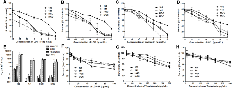
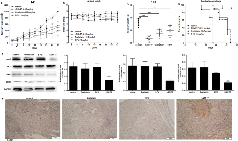
Circulating tumor cells (CTCs) are a major cause of tumor metastasis and resistance to anticancer therapies. To date, no effective low-toxicity chemotherapeutic agents or antibodies have exhibited significant clinical activity against CTCs. Macrophages are important mediators of antitumor immunity. Tuftsin (TF), a tetrapeptide located at residues 289-292 of the CH2 domain of the Fc region of the IgG heavy chain, binds to Nrp-1, a receptor on the surface of macrophages that promotes phagocytosis and induces nonspecific activation of the immune system against tumors. Lidamycin (LDM) is an antitumor chemotherapy agent that is strongly cytotoxic to tumors and can dissociate into an apoprotein (LDP) and active enediyne (AE) in vitro. We previously constructed the fusion protein LDP-TF through genetic engineering and inserted the chromophore AE to produce LDM-TF, which can target macrophages to promote their phagocytic and cytotoxic activity against tumor cells. Preliminary experiments confirmed the anti-tumor activity of LDM-TFs. In this study, we found that LDM-TF effectively inhibited the growth of CTCs of gastric cancer origin and enhanced macrophage phagocytosis both in vivo and in vitro. Tumor cell expression of CD47, which helps to evade phagocytosis by macrophages, was substantially downregulated by LDM-TF. Notably, our in vitro experiments demonstrated that the combination of LDM-TF and anti-CD47 antibodies promoted phagocytosis more than either component alone. Our findings demonstrate the significant inhibitory effect of LDM-TF on the growth of CTCs of gastric cancer origin and suggest that the combination of LDM-TF and anti-CD47 antibodies may exhibit synergistic effects, thereby providing a new option for the clinical treatment of patients with advanced tumors that have metastasized.
Keywords: CD47; Circulating tumor cells; Fusion protein; Lidamycin; Macrophage; Tuftsin.
© 2023 The Authors.
Conflict of interest statement
The authors declare that they have no known competing financial interests or personal relationships that could have appeared to influence the work reported in this paper.
Figures


Similar articles
-
Tuftsin-based, EGFR-targeting fusion protein and its enediyne-energized analog show high antitumor efficacy associated with CD47 down-regulation.Cancer Immunol Immunother. 2014 Dec;63(12):1261-72. doi: 10.1007/s00262-014-1604-1. Epub 2014 Aug 28. Cancer Immunol Immunother. 2014. PMID: 25164878 Free PMC article.
-
A novel enediyne-integrated antibody-drug conjugate shows promising antitumor efficacy against CD30+ lymphomas.Mol Oncol. 2018 Mar;12(3):339-355. doi: 10.1002/1878-0261.12166. Epub 2018 Jan 26. Mol Oncol. 2018. PMID: 29316337 Free PMC article.
-
An EGFR/CD13 bispecific fusion protein and its enediyne-energized analog show potent antitumor activity.Anticancer Drugs. 2014 Jan;25(1):82-91. doi: 10.1097/CAD.0000000000000029. Anticancer Drugs. 2014. PMID: 24100279
-
Macrophages are critical effectors of antibody therapies for cancer.MAbs. 2015;7(2):303-10. doi: 10.1080/19420862.2015.1011450. MAbs. 2015. PMID: 25667985 Free PMC article. Review.
-
Phagocytic function of tumor-associated macrophages as a key determinant of tumor progression control: a review.J Immunother Cancer. 2020 Dec;8(2):e001408. doi: 10.1136/jitc-2020-001408. J Immunother Cancer. 2020. PMID: 33335026 Free PMC article. Review.
Cited by
-
Functionalizing Dendrimers for Targeted Delivery of Bioactive Molecules to Macrophages: A Potential Treatment for Mycobacterium tuberculosis Infection-A Review.Pharmaceuticals (Basel). 2023 Oct 9;16(10):1428. doi: 10.3390/ph16101428. Pharmaceuticals (Basel). 2023. PMID: 37895899 Free PMC article. Review.
References
-
- Liu W.J., Liu X.J., Li L., et al. Tuftsin-based, EGFR-targeting fusion protein and its enediyne-energized analog show high antitumor efficacy associated with CD47 down-regulation, CANCER IMMUNOL IMMUN. Cancer Immunol. Immunother. 2014;63:1261–1272. doi: 10.1007/s00262-014-1604-1. - DOI - PMC - PubMed
LinkOut - more resources
Full Text Sources
Research Materials
Miscellaneous
