. 2023 Jan 22;5(3):114-130.
doi: 10.1096/fba.2022-00098. eCollection 2023 Mar.

Analyzing the interactome of human CK2β in prostate carcinoma cells reveals HSP70-1 and Rho guanin nucleotide exchange factor 12 as novel interaction partners

Affiliations

Analyzing the interactome of human CK2β in prostate carcinoma cells reveals HSP70-1 and Rho guanin nucleotide exchange factor 12 as novel interaction partners

Anna Nickelsen et al. FASEB Bioadv. .

Abstract

CK2β is the non-catalytic modulating part of the S/T-protein kinase CK2. However, the overall function of CK2β is poorly understood. Here, we report on the identification of 38 new interaction partners of the human CK2β from lysates of DU145 prostate cancer cells using photo-crosslinking and mass spectrometry, whereby HSP70-1 was identified with high abundance. The KD value of its interaction with CK2β was determined as 0.57 μM by microscale thermophoresis, this being the first time, to our knowledge, that a KD value of CK2β with another protein than CK2α or CK2α' was quantified. Phosphorylation studies excluded HSP70-1 as a substrate or activity modulator of CK2, suggesting a CK2 activity independent interaction of HSP70-1 with CK2β. Co-immunoprecipitation experiments in three different cancer cell lines confirmed the interaction of HSP70-1 with CK2β in vivo. A second identified CK2β interaction partner was Rho guanin nucleotide exchange factor 12, indicating an involvement of CK2β in the Rho-GTPase signal pathway, described here for the first time to our knowledge. This points to a role of CK2β in the interaction network affecting the organization of the cytoskeleton.

Keywords: HSP70‐1; Rho guanin nucleotide exchange factor 12; photo‐crosslinking mass spectrometry; prostate cancer; protein kinase CK2.


Figures

FIGURE 1
FIGURE 1
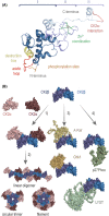
Schematic structure of CK2β as well as interaction sites and interaction partners. (A) Structure of human CK2β monomer (PDB: 1jwh) sectioned into (I) N‐terminal region (E5‐G104), (II) juxta dimer interface (D105‐T161) and (III) C‐terminal region (G162‐R215). Functional areas were highlighted: phosphorylation sites (S2, S3; orange), destruction box (R47‐D54; yellow), acidic loop (D55‐P66; red), zinc coordinating amino acids (C109, C114, C137 and C140; green), main interaction surface with CK2α (N181‐A203; light red). (B) Known interactions of the CK2β dimer. (1) Binding of CK2 catalytic subunit CK2α assembling the tetrameric CK2 holoenzyme. (2) Aggregation of several CK2 holoenzymes. (3) interaction with other kinases shown as a model, A‐Raf (AF‐P10398‐F1‐mod), Chk1 (AF‐014757‐F1‐mod), (4) interaction with regulatory proteins shown as a model, p27Phox/NCF1 (AF‐P14598‐F1‐mod), LYST (AF‐E9QFZ1‐F1‐mod).
FIGURE 2
FIGURE 2
Incorporation sites of pAzF in CK2β and enzyme activity of CK2 holoenzymes containing the different variants in comparison to unmodified CK2β. (A) CK2 holoenzyme consisting of two CK2α and two CK2β subunits (PDB: 1jwh) and the CK2β variants with pAzF at position 34 (yellow), 62 (petrol) and 80 (red). (B) Phosphorylation of CK2 substrate peptide RRRDDDSDDD (300 μM) by CK2α or CK2α based holoenzymes was analyzed using the capillary electrophoresis‐based activity assay. CK2α (40 nM) was incubated with 80 nM CK2β or modified variants CK2β34pAzF, CK2β62pAzF or CK2β80pAzF at 37°C. Mean ± SD, n = 3, *: p ≤ 0.05, n.s.: p > 0.05.
FIGURE 3
FIGURE 3
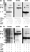
UV light induced crosslinking of CK2β34pAzF with CK2α. (A) CK2β34pAzF (10 μM) and CK2α (3 μM) were incubated for 15 min at 37°C, UV light irradiated (λ = 365 nm) and purified by NiNTA affinity chromatography. (B) CK2β34pAzF (10 μM) and CK2α (3 μM) were incubated in DU145 cell lysate (5 mg/ml protein), UV light irradiated and purified by NiNTA affinity chromatography. Proteins were separated by SDS‐PAGE and analyzed by Coomassie staining or western blot with primary antibody against CK2α or against 6xHis‐tag.
FIGURE 4
FIGURE 4
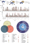
Identification and analysis of CK2β interaction partners. (A) Experimental workflow. One of the CK2β variants (20 μM) or no external protein as control were mixed each with lysate of DU145 cell lysate, photo‐crosslinking was performed by UV light irradiation. Cross‐linked products were enriched by NiNTA affinity chromatography followed by harsh washing conditions. After tryptic digestion, proteins coupled to CK2β were analyzed using LC–MS. (B) Ion intensities of the proteins identified in samples containing CK2β34pAzF (yellow), CK2β62pAzF (petrol) or CK2β80pAzF (red) were related to the intensity of the corresponding CK2β variant (IβV) or (C) to the intensity of the respective protein in the control (IC). Proteins already described as interacting partner of CK2β (filled circles) or CK2α (unfilled circles) as well as proteins showing at least for one variant a mean IβV > 0.01 (triangles) or a mean IC > 100 (squares) were marked. Experiment was performed in two independent replicates and data are shown as mean including range. Proteins that were not detected in control in at least one replicate were marked with a gray diamond. (D) Distribution of proteins (P) on the respective variant in which presence they were found with an IβV value at least 2.5‐fold higher than in the presence of the other variants. These proteins were also marked in the list with a diamond in the respective color. (E) Assignment of identified proteins to their physiological function.
FIGURE 5
FIGURE 5
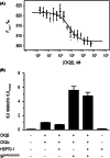
Characterization of the interaction of CK2β and HSP70‐1. (A) The binding affinity of fluorescently labeled HSP70‐1 to CK2β was measured by microscale thermophoresis. The KD‐value was determined to 0.57 μM. Data is shown as mean ± SD, n = 4. (B) Phosphorylation through CK2α supplemented with CK2β in the ratio 1:2 was analyzed in presence or absence of 1 μM HSP70‐1 and 300 μM of the CK2 substrate peptide (SP) RRRDDDSDDD by ADP‐Glo™ assay. The rLU were related to the mean rLU value of the samples containing only CK2α and CK2β. Data are shown as mean ± SD, n = 3.
FIGURE 6
FIGURE 6
Co‐immunoprecipitation of CK2β and HSP70 in extracts of DU145, LNCaP and HeLa cells. Cells were harvested after or before heat shock at 42°C for 30 min. Cell extracts were prepared, and an amount corresponding to 1 mg protein was incubated with pure protein G sepharose beads (pure beads). After removing pure beads, cell extracts were incubated with beads carrying an CK2β serum for co‐immunoprecipitation (β‐serum beads). Precipitates were separated by SDS‐PAGE and analyzed by western blot subsequently using a primary antibody against CK2β first, a primary antibody against HSP70 and finally a primary antibody against CK2α. Extracts of all cell lines were incubated with mere protein G sepharose beads prior to co‐immunoprecipitation to remove proteins binding non‐specifically. IgG HC: IgG heavy chains.
FIGURE 7
FIGURE 7
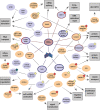
Interactome of the 43 CK2β binding proteins identified in this study with regard to their physiological function. Protein interactions listed on BioGrid are marked by lines. Proteins already known to interact with CK2β are framed in blue, proteins that have a homolog known to interact with CK2β are highlighted in blue. Known interaction partners of CK2α are framed in red. Proteins that have not been listed as interaction partners of CK2β or CK2α before or that belong to protein families that are known to interact with CK2β are colored in orange. A rough classification according to the physiological function of the proteins as presented is indicated by arrows. Proteins found with a 2.5‐fold excess in the presence of one of the three variants compared to the others are marked with a colored diamond: CK2β34pAzF (yellow), CK2β62pAzF (petrol) and CK2β80pAzF (red).
FIGURE 8
FIGURE 8
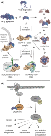
Hypothetical model of possible interaction pathways of CK2β with HSP70‐1 and the influence of CK2β‐LARG interaction on RhoA signaling. (A) The interaction of CK2β (PDB 1jwh) with HSP70‐1 (PDB 3jxu—Nucleotide binding domain, PDB 4wv5—Substrate binding domain) may affect CK2β or CK2 regulation, various ongoing processes through interaction in heterogenic multicomplexes with clathrin (PDB 3lvh) and HERC1 or with HSP90 (PDB 2iop) as well as it may affect the regulation of lysosomes. (B) LARG is activated by G‐protein coupled receptor activated G‐protein Gα13, and normally activates RhoA responsible for migration and invasion of prostate cancer cells as well as for cytoskeleton reorganization, stress fiber formation and actin polymerization. The function of CK2β binding to LARG thereon is still unclear.

Similar articles

Cited by

References

    1. Heller‐Harrison RA, Meisner H, Czech MP. Cloning and characterization of a cDNA encoding the beta subunit of human casein kinase II. Biochemistry. 1989;28:9053‐9058. - PubMed
    1. Jakobi R, Voss H, Pyerin W. Human phosvitin/casein kinase type II. Molecular cloning and sequencing of full‐length cDNA encoding subunit beta. Eur J Biochem. 1989;183:227‐233. - PubMed
    1. Boldyreff B, Piontek K, Schmidt‐Spaniol I, Issinger OG. The beta subunit of casein kinase II: cloning of cDNAs from murine and porcine origin and expression of the porcine sequence as a fusion protein. Biochim Biophys Acta. 1991;1088:439‐441. - PubMed
    1. Livak KJ. Detailed structure of the Drosophila melanogaster stellate genes and their transcripts. Genetics. 1990;124:303‐316. - PMC - PubMed
    1. Egorova KS, Olenkina OM, Kibanov MV, Kalmykova AI, Gvozdev VA, Olenina LV. Genetically derepressed nucleoplasmic stellate protein in spermatocytes of D. melanogaster interacts with the catalytic subunit of protein kinase 2 and carries histone‐like lysine‐methylated mark. J Mol Biol. 2009;389:895‐906. - PubMed

LinkOut - more resources