. 2023 Mar 6;11(2):e0397422.
doi: 10.1128/spectrum.03974-22. Online ahead of print.

Proteomic Study of the Interactions between Phages and the Bacterial Host Klebsiella pneumoniae

Affiliations

Proteomic Study of the Interactions between Phages and the Bacterial Host Klebsiella pneumoniae

Inés Bleriot et al. Microbiol Spectr. .

Abstract

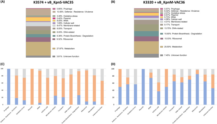
Phages and bacteria have acquired resistance mechanisms for protection. In this context, the aims of the present study were to analyze the proteins isolated from 21 novel lytic phages of Klebsiella pneumoniae in search of defense mechanisms against bacteria and also to determine the infective capacity of the phages. A proteomic study was also conducted to investigate the defense mechanisms of two clinical isolates of K. pneumoniae infected by phages. For this purpose, the 21 lytic phages were sequenced and de novo assembled. The host range was determined in a collection of 47 clinical isolates of K. pneumoniae, revealing the variable infective capacity of the phages. Genome sequencing showed that all of the phages were lytic phages belonging to the order Caudovirales. Phage sequence analysis revealed that the proteins were organized in functional modules within the genome. Although most of the proteins have unknown functions, multiple proteins were associated with defense mechanisms against bacteria, including the restriction-modification system, the toxin-antitoxin system, evasion of DNA degradation, blocking of host restriction and modification, the orphan CRISPR-Cas system, and the anti-CRISPR system. Proteomic study of the phage-host interactions (i.e., between isolates K3574 and K3320, which have intact CRISPR-Cas systems, and phages vB_KpnS-VAC35 and vB_KpnM-VAC36, respectively) revealed the presence of several defense mechanisms against phage infection (prophage, defense/virulence/resistance, oxidative stress and plasmid proteins) in the bacteria, and of the Acr candidate (anti-CRISPR protein) in the phages. IMPORTANCE Researchers, including microbiologists and infectious disease specialists, require more knowledge about the interactions between phages and their bacterial hosts and about their defense mechanisms. In this study, we analyzed the molecular mechanisms of viral and bacterial defense in phages infecting clinical isolates of K. pneumoniae. Viral defense mechanisms included restriction-modification system evasion, the toxin-antitoxin (TA) system, DNA degradation evasion, blocking of host restriction and modification, and resistance to the abortive infection system, anti-CRISPR and CRISPR-Cas systems. Regarding bacterial defense mechanisms, proteomic analysis revealed expression of proteins involved in the prophage (FtsH protease modulator), plasmid (cupin phosphomannose isomerase protein), defense/virulence/resistance (porins, efflux pumps, lipopolysaccharide, pilus elements, quorum network proteins, TA systems, and methyltransferases), oxidative stress mechanisms, and Acr candidates (anti-CRISPR protein). The findings reveal some important molecular mechanisms involved in the phage-host bacterial interactions; however, further study in this field is required to improve the efficacy of phage therapy.

Keywords: Klebsiella; Klebsiella pneumoniae; bacteriophage; bacteriophage evolution; defense mechanism; lytic phage; phage-host interaction; plasmid; prophage; virus-host interactions.


Conflict of interest statement

The authors declare no conflict of interest.

Figures

FIG 1
FIG 1
(A) Phylogenetic analysis of 21 phages performed with the nucleotide sequence of the large terminase subunit of each phage. (B) TEM images showing the structure of the 21 phages under study. All belong to the order Caudovirales. vB_KpnS-VAC2, vB_KpnS-VAC4, vB_KpnS-VAC5, vB_KpnS-VAC6, vB_KpnS-VAC7, vB_KpnS-VAC8, vB_KpnS-VAC10, vB_KpnS-VAC11, vB_KpnS-VAC35, vB_KpnS-VAC70, vB_KpnS-VAC110, vB_KpnS-VAC111, vB_KpnS-VAC112, and vB_KpnS-VAC113 are characterized by large and flexible tails. On the other hand, phages vB_KpnP-VAC1, vB_KpnP-VAC25, and vB_KpnP-VAC71 are characterized by a short tail. Finally, phages vB_KpnM-VAC13, vB_KpnM-VAC36, and vB_KpnM-VAC66 are characterized by an icosahedral capsid and a rigid, contractile tail. The TEM scale bar represents 50 or 100 nm, depending on the phage.
FIG 2
FIG 2
Graphic comparison of the homology of the 21 phages, grouped according to their families and in the same order as in the phylogenetic tree. The schematic representation was conducted with VipTree (https://www.genome.jp/viptree/, accessed in June 2022).
FIG 3
FIG 3
(A) Schematic representation of the host range technique. (B) Host range of the 21 phages included in the collection of 47 clinical strains of K. pneumoniae and the percentage of infectivity. The strains indicated in red are strains were infected by vB_KpnS-VAC35 and vB_KpnM-VAC36 phages, and an asterisk (*) represents the presence of the CRISPR-Cas system within these strains.
FIG 4
FIG 4
(A) Scheme of the modular organization of class I, type I CRISPR-Cas systems. (Diagram adapted from reference .) SS* indicates the putative small subunit (SS) that might be fused to the large subunit in several type I subtypes. (B and C) Graphic representation of the CRISPR-Cas system of strains K3574 and K3320, and adapted image of CRISPR Miner 2 (http://www.microbiome-bigdata.com/CRISPRminer, accessed in March 2022). The red square represents the repeats. Whereas the black and gray diamonds represent spacer sequences, the numbers below the repeats represent the numbers of spacers. The arrows represent the open reading frames, and the different colors represent the different Cas types.
FIG 5
FIG 5
(A and B) Graphic representation of the genome of the phage vB_KpnS-VAC35 and vB_KpnM-VAC36, constructed with the Snapgene tool, v6.0.5. (C and D) Adsorption curve of phages vB_KpnS-VAC35 and vB_KpnM-VAC36, with adsorption times of 5 and 2 min, respectively. The error bars represent the standard deviations of three experimental replicates. (E and F) One-step growth curve of phages vB_KpnS-VAC35 and vB_KpnM-VAC36, with latent times (labeled “L”) of 10 and 8 min and a burst size (labeled “B”) of 45.52 PFU/mL and 2.71, respectively. The error bars represent the standard deviations of three experimental replicates.
FIG 6
FIG 6
(A and B) Infection curve of the strains K3574 (orange) and K3320 (blue), respectively, with phages vB_KpnS-VAC35 (light orange) and vB_KpnM-VAC36 (light blue) at an MOI of 1. (C and D) Measurement of viability by CFU/mL counts of strains K3574 and K3320 infected with, respectively, phage vB_KpnS-VAC35 and vB_KpnM-VAC36, at an MOI of 1 over time. (E and F) Measurement of PFU/mL counts of the phages vB_KpnS-VAC35 and vB_KpnM-VAC36 at an MOI 1 over time.
FIG 7
FIG 7
(A and B) Graphical representation of the proteomics results, showing the abundance of each group of proteins found in the culture with the bacterial strain K3574 infected with phage vB_KpnS-VAC35 and in the culture with the bacterial strain K3320 infected with phage vB_KpnM-VAC36. (C and D) Abundance of proteins with a higher (blue), lower (orange), or undetected (gray) value areas compared to the uninfected control in strains K3574 and K3320 infected with phages vB_KpnS-VAC35 and vB_KpnM-VAC36, respectively.

References

    1. Bergh O, Børsheim KY, Bratbak G, Heldal M. 1989. High abundance of viruses found in aquatic environments. Nature 340:467–468. doi:10.1038/340467a0. - DOI - PubMed
    1. Bleriot I, Trastoy R, Blasco L, Fernández-Cuenca F, Ambroa A, Fernández-García L, et al.. 2020. Genomic analysis of 40 prophages located in the genomes of 16 carbapenemase-producing clinical strains of Klebsiella pneumoniae. Microb Genom 6. doi:10.1099/mgen.0.000369. - DOI - PMC - PubMed
    1. Domingo-Calap P, Delgado-Martínez J. 2018. Bacteriophages: protagonists of a post-antibiotic era. Antibiotics (Basel) 7. doi:10.3390/antibiotics7030066. - DOI - PMC - PubMed
    1. Taati Moghadam M, Khoshbayan A, Chegini Z, Farahani I, Shariati A. 2020. Bacteriophages, a new therapeutic solution for inhibiting multidrug-resistant bacteria causing wound infection: lesson from animal models and clinical trials. Drug Des Devel Ther 14:1867–1883. doi:10.2147/DDDT.S251171. - DOI - PMC - PubMed
    1. Furfaro LL, Payne MS, Chang BJ. 2018. Bacteriophage therapy: clinical trials and regulatory hurdles. Front Cell Infect Microbiol 8:376. doi:10.3389/fcimb.2018.00376. - DOI - PMC - PubMed

LinkOut - more resources