. 2023 Nov-Dec;25(6):478-486.
doi: 10.1089/fpsam.2022.0346. Epub 2023 Mar 6.

Biodegradable Nerve Guide with Glial Cell Line-Derived Neurotrophic Factor Improves Recovery After Facial Nerve Injury in Rats

Affiliations

Biodegradable Nerve Guide with Glial Cell Line-Derived Neurotrophic Factor Improves Recovery After Facial Nerve Injury in Rats

Fuat Baris Bengur et al. Facial Plast Surg Aesthet Med. 2023 Nov-Dec.

Abstract

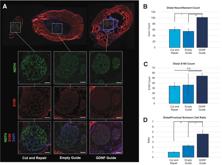
Background: Bioengineered nerve guides with glial cell line-derived neurotrophic factor (GDNF) support recovery after facial nerve injury by acting as regenerative scaffolds. Objective: To compare functional, electrophysiological, and histological outcomes after repair of rat facial nerve transection in control, empty nerve guide, and nerve guide with GDNF conditions. Methods: Rats underwent transection and primary repair of the buccal branch of the facial nerve and were divided into (1) transection and repair only, (2) transection and repair augmented with empty guide, (3) transection and repair augmented with GDNF-guide groups. Weekly measurements of the whisking movements were recorded. At 12 weeks, compound muscle action potentials (CMAPs) at the whisker pad were assessed, and samples were collected for histomorphometric analysis. Results: Rats in GDNF-guide group displayed the earliest peak in normalized whisking amplitude. CMAPs were significantly higher after GDNF-guide placement. Mean fiber surface area of the target muscle, axonal count of the injured branch, and the number of Schwann cells were highest with GDNF guides. Conclusion: The biodegradable nerve guide containing double-walled GDNF microspheres enhanced recovery after facial nerve transection and primary repair.


Conflict of interest statement

No competing financial interests exist.

Figures

Fig. 1.
Fig. 1.
The surgical model. (A) The inferior border of the mandible was marked for a 1.5 cm curvilinear incision. (B) After dissection of the subcutaneous tissues, the buccal and marginal mandibular branches were identified. The buccal branch was cut and primarily repaired (star), while a 5 mm segment was resected from the marginal mandibular branch and both ends were ligated with sutures (arrows). (C) The nerve guide was placed around the repaired buccal branch and secured with a suture (star) to the underlying fascia. (D–F) The repaired nerves in all experimental conditions at the 12-week endpoint: (D) transection and repair only, (E) transection and repair with empty nerve guide, and (F) transection and repair with GDNF-containing guide. Scale bars: 1 cm. GDNF, glial cell line-derived neurotrophic factor.
Fig. 2.
Fig. 2.
The whisker movement measurements (n = 5 for each group). (A) On still images, a sagittal midline was determined by extending a perpendicular line from the middle point of the medial angle of both eyes toward the nose. The rostrally open angle of the marked whiskers with the midline point was determined by selecting the base of the whiskers and the black marking about 1 cm distal on the whiskers. Representative measurements of the protraction and retraction angles on both sides at the baseline can be visualized. Arrows indicate the C1 whiskers on each side that were marked with a black marker, prior to the measurements. The contrast of the black color with the whiskers allowed for accurate quantification of the movements. (B) The percent recovery of the whisking amplitude from the initial week after the surgery to 6- and 12-week timepoints. A significant recovery was achieved in all groups after the surgery, while only the GDNF-guide group showed significant improvement at the 6-week timepoint. (C) The longitudinal recovery of both groups with two-point moving average. GDNF-guide group had the earliest peak and highest overall recovery. Error bars represent standard deviation. *p < 0.05, **p < 0.01, ***p < 0.001. ΔOne animal was not able to complete the whisking measurements. n.s., not significant.
Fig. 3.
Fig. 3.
CMAP measurements (n = 5 for each group). (A) At the endpoint, the buccal branch was exposed, and the custom-built electrode cuffs were placed distal to the nerve guide (star). Subdermal needle electrodes were placed into the vibrissal muscles on rows C and D. Scale bar: 1 cm. (B) Average representative waveforms of CMAP recorded from the whisker pads of rats in all experimental conditions. (C) The mean peak-to-peak amplitude of CMAP in all experimental conditions. Error bars represent standard error. ***p < 0.001. CMAP, compound muscle action potential.
Fig. 4.
Fig. 4.
Muscle fiber count analysis (n = 5 for each group). (A) Levator labii superioris muscles in the denervated sides in all three experimental conditions were stained with Gardner's Trichrome (top) and H&E (bottom). Note the prominent atrophy of the muscle fibers on the cut and repair and empty guide groups. Scale bar: 100 μm. (B) Regions of interest in each muscle were analyzed to identify the mean surface area of the fibers. GDNF-treated group had significantly higher mean muscle fiber diameter. Error bars represent standard error. *p < 0.05, ***p < 0.001. H&E, hematoxylin and eosin.
Fig. 5.
Fig. 5.
Immunofluorescence staining of the nerve samples (n = 3 for each group). (A) Samples were stained for NEFH (green) for axons, S100 (red) for Schwann cells, and DAPI (blue) for nuclei. Scale bars: 100 μm. (B) The GDNF treated group showed the highest axonal count versus others on the region distal to the injury. (C) The GDNF treated group showed the highest Schwann cell count on the region distal to the injury; however, the difference failed to reach statistical significance. (D) Compared with the proximal segments, the ratio of Schwann cells was significantly higher in the area distal to the injury in the GDNF-treated group. Error bars represent standard error. *p < 0.05, **p < 0.01, ***p < 0.001. DAPI, 4′,6-diamidino-2-phenylindole; NEFH, neurofilament heavy chain.

References

    1. Bradbury ET, Simons W, Sanders R. Psychological and social factors in reconstructive surgery for hemi-facial palsy. J Plast Reconstruct Aesthet Surg. 2006;59(3):272–278. - PubMed
    1. Coulson SE, O'Dwyer NJ, Adams RD, Croxson GR. Expression of emotion and quality of life after facial nerve paralysis. Otol Neurotol. 2004;25(6):1014–1019. - PubMed
    1. Hohman MH, Hadlock TA. Etiology, diagnosis, and management of facial palsy: 2000 patients at a facial nerve center. Laryngoscope. 2014;124(7). doi:10.1002/lary.24542 - DOI - PubMed
    1. Gordin E, Lee TS, Ducic Y, Arnaoutakis D. Facial nerve trauma: evaluation and considerations in management. Craniomaxillofac Trauma Reconstr. 2015;8(1):1–13. - PMC - PubMed
    1. Lorch M, Teach SJ. Etiology and approach to diagnosis and treatment. Pediatr Emerg Care. 2010;26(10):10. - PubMed

Substances

LinkOut - more resources