. 2023 Mar 15;145(10):5824-5833.
doi: 10.1021/jacs.2c13049. Epub 2023 Mar 6.

Triggered Polymersome Fusion

Affiliations

Triggered Polymersome Fusion

Stephen D P Fielden et al. J Am Chem Soc. .

Abstract

The contents of biological cells are retained within compartments formed of phospholipid membranes. The movement of material within and between cells is often mediated by the fusion of phospholipid membranes, which allows mixing of contents or excretion of material into the surrounding environment. Biological membrane fusion is a highly regulated process that is catalyzed by proteins and often triggered by cellular signaling. In contrast, the controlled fusion of polymer-based membranes is largely unexplored, despite the potential application of this process in nanomedicine, smart materials, and reagent trafficking. Here, we demonstrate triggered polymersome fusion. Out-of-equilibrium polymersomes were formed by ring-opening metathesis polymerization-induced self-assembly and persist until a specific chemical signal (pH change) triggers their fusion. Characterization of polymersomes was performed by a variety of techniques, including dynamic light scattering, dry-state/cryogenic-transmission electron microscopy, and small-angle X-ray scattering (SAXS). The fusion process was followed by time-resolved SAXS analysis. Developing elementary methods of communication between polymersomes, such as fusion, will prove essential for emulating life-like behaviors in synthetic nanotechnology.


Conflict of interest statement

The authors declare no competing financial interest.

Figures

Figure 1
Figure 1
(a) Triggered biological membrane fusion proceeds over two steps. First, membranes are brought into close contact by the complementary interactions of SNARE proteins to generate a metastable state (with respect to the final fused product). Next, an influx of Ca2+ promotes protein rearrangement, which triggers fusion of the two membranes. (b) In this work, polymersomes are produced in a persistent metastable state. They then undergo rapid fusion on application of a trigger. Relative thicknesses of layers in polymersome cartoons are not to scale.
Figure 2
Figure 2
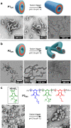
TEM analysis of P1x and P2x nano-object products when NB-MEG DP = 200 (unfused) or 300 (fused). P1x particles contain a corona formed of a diblock copolymer (blue and green layers in cartoon). P2x particles contain a corona formed of a random copolymer (aquamarine layer in cartoon). Hydrophobic layers in cartoons are red. Dry-state TEM samples were stained with 1% uranyl acetate solution prior to imaging.
Figure 3
Figure 3
Triggered polymersome fusion by a pH switch. Continuous phase at pH 2: 90 vol % 100 mM PB2 + 10 vol % THF. Continuous phase at pH 12: 90 vol % (25 mM PB2 + 75 mM NaOH) + 10 vol % THF. TEM analysis (dry-state and cryo-) of (a) fused P1200 particles and (b) fused and aggregated P2200 particles. (c) Structure and attempted fusion of P3200, which contains pH unresponsive P(NB-NR4) rather than P(NB-amine). Dry-state TEM samples were stained with 1% uranyl acetate solution prior to imaging.
Figure 4
Figure 4
Mechanistic analysis of fusion of P1200 particles by SAXS. (a) Static SAXS data of unfused P1200 particles fitted to a spherical micelle model (red line). (b) Static SAXS data of fused P1200 particles fitted to a cylindrical micelle model (red line). (c) Evolution of cylinder length over time during in situ SAXS analysis. The dashed gray line corresponds to the cylinder length from modeling of the static measurement in part (b). Line added to guide the eye. (d) Evolution of cylinder width over time during in situ SAXS analysis. The dashed gray line corresponds to the cylinder width from modeling of the static measurement in part (b). Line added to guide the eye. (e) Evolution in volume fraction of spheres [unfused particles] and cylinders [fused particles] during in situ SAXS analysis. Values obtained from weighting of models to give the best fit. (f) Proposed two-step fusion mechanism with dry-state TEM images of particles quenched after 5 s and 5 min at pH 12. Dry-state TEM samples were stained with 1% uranyl acetate solution prior to imaging.
Figure 5
Figure 5
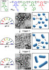
Multistep triggered fusion with tetrablock copolymer P4200 containing two pH-responsive blocks. The first trigger (switching pH from 2 to 7) deprotonates the P(NB-Py) block, resulting in partial fusion. The second trigger deprotonates the P(NB-amine) block, resulting in complete fusion. Dry-state TEM samples were stained with 1% uranyl acetate solution prior to imaging.

References

    1. Diekmann Y.; Pereira-Leal J. B. Evolution of intracellular compartmentalization. Biochem. J. 2013, 449, 319–331. 10.1042/BJ20120957. - DOI - PubMed
    1. Marguet M.; Bonduelle C.; Lecommandoux S. Multicompartmentalized polymeric systems: towards biomimetic cellular structure and function. Chem. Soc. Rev. 2013, 42, 512–529. 10.1039/C2CS35312A. - DOI - PubMed
    1. Howell G. J.; Holloway Z. G.; Cobbold C.; Monaco A. P.; Ponnambalam S. Cell Biology of Membrane Trafficking in Human Disease. Int. Rev. Cytol. 2006, 252, 1–69. 10.1016/S0074-7696(06)52005-4. - DOI - PMC - PubMed
    1. Yarwood R.; Hellicar J.; Woodman P. G.; Lowe M. Membrane trafficking in health and disease. Dis. Model Mech. 2020, 13, dmm043448.10.1242/dmm.043448. - DOI - PMC - PubMed
    1. Lu T.; Guo H. How the Membranes Fuse: From Spontaneous to Induced. Adv. Theory Simul. 2019, 2, 1900032.10.1002/adts.201900032. - DOI

Publication types