. 2023 Mar 14;120(11):e2218247120.
doi: 10.1073/pnas.2218247120. Epub 2023 Mar 6.

Biolistic delivery of liposomes protected in metal-organic frameworks

Affiliations

Biolistic delivery of liposomes protected in metal-organic frameworks

Sneha Kumari et al. Proc Natl Acad Sci U S A. .

Abstract

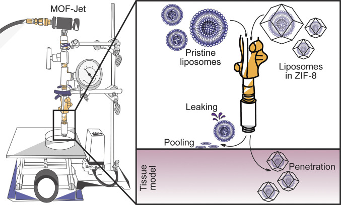
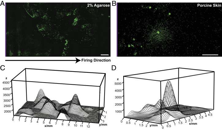
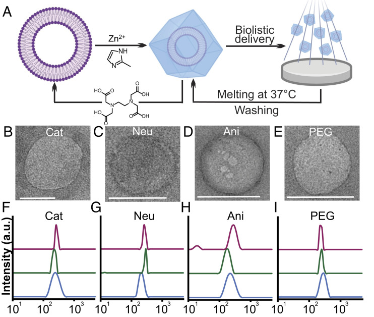
Needle-and-syringe-based delivery has been the commercial standard for vaccine administration to date. With worsening medical personnel availability, increasing biohazard waste production, and the possibility of cross-contamination, we explore the possibility of biolistic delivery as an alternate skin-based delivery route. Delicate formulations like liposomes are inherently unsuitable for this delivery model as they are fragile biomaterials incapable of withstanding shear stress and are exceedingly difficult to formulate as a lyophilized powder for room temperature storage. Here we have developed a approach to deliver liposomes into the skin biolistically-by encapsulating them in a nano-sized shell made of Zeolitic Imidazolate Framework-8 (ZIF-8). When encapsulated within a crystalline and rigid coating, the liposomes are not only protected from thermal stress, but also shear stress. This protection from stressors is crucial, especially for formulations with cargo encapsulated inside the lumen of the liposomes. Moreover, the coating provides the liposomes with a solid exterior that allows the particles to penetrate the skin effectively. In this work, we explored the mechanical protection ZIF-8 provides to liposomes as a preliminary investigation for using biolistic delivery as an alternative to syringe-and-needle-based delivery of vaccines. We demonstrated that liposomes with a variety of surface charges could be coated with ZIF-8 using the right conditions, and this coating can be just as easily removed-without causing any damage to the protected material. The protective coating prevented the liposomes from leaking cargo and helped in their effective penetration when delivered into the agarose tissue model and porcine skin tissue.

Keywords: biolistic delivery; biomimetic mineralization; metal-organic frameworks; shear stress; zeolitic imidazolate frameworks.


Conflict of interest statement

The authors declare no competing interest.

Figures

Fig. 1.
Fig. 1.
Comparative studies between pristine and ZIF-8-coated liposomes demonstrate the mechanical stability of the ZIF-8 coating. Pristine liposomes leak under the shear stress of the MOF-Jet, while ZIF-8-coated liposomes remain protected and negligible leaking is observed. The hard coating of the ZIF-8 allows the coated liposomes to penetrate into the tissue model, while the pristine liposomes just pool on the surface.
Fig. 2.
Fig. 2.
(AD) TEM micrographs of pristine Cat, Neu, Ani, and PEG liposomes. (Scale bar, 200 nm for all micrographs.) (E) Fluorescence spectrum of Cy5@Neu. (F) Pellet of Cy5@Neu obtained after ultracentrifugation at 160,000× g and removal of excess Cy5 dye in the supernatant. No significant differences were observed between the fluorescence spectra and pellets of Cy5@Neu, Cy5@Cat, Cy5@Ani, and Cy5@PEG. (G) ζ-potential of pristine Cat, Neu, Ani, and PEG liposomes.
Fig. 3.
Fig. 3.
(AD) Encapsulation efficiencies of Cy5@Cat, Cy5@Neu, Cy5@Ani, and Cy5@PEG liposomes at various L/M ratios. Each of the liposomes was encapsulated in ZIF-8 by adding ZnOAc and HMIM such that the L/M ratios were 32, 16, 8, and 4—as labeled on the x axis. All conditions were measured for encapsulation efficiencies after 3 h (orange) and 24 h (yellow). Measurements were performed by quantifying the percentage of unencapsulated Cy5@Lip in the supernatant using fluorescence. Error bars are represented as ±SD. (E) PXRD pattern of Neu@Z-32 and simulated ZIF-8. (F) SEM micrograph of Neu@Z-32. (Scale bar, 1 µm.) (G) Epifluorescence micrograph of Neu@Z-32. (Scale bar, 100 µm.) No significant differences were observed in the PXRD patterns, SEM micrographs, and epifluorescence micrographs of Neu@Z-32, Cat@Z-32, Ani@Z-32, PEG@Z-32, Neu@Z-16, Cat@Z-16, Ani@Z-16, and PEG@Z-16.
Fig. 4.
Fig. 4.
(A) Schematic illustration of AFM nano-indentation of ZIF-8 and Lip@Z crystals. (B) Image of Lip@Z crystal captured before and after nano-indentation. (Scale bar, 100 nm for both images.) (C) Single representative force-displacement curves of free ZIF-8 control and Lip@Z, respectively. (D) Fracture force of ZIF-8 and Lip@Z measured from the force-displacement curves (n = 5 for each).
Fig. 5.
Fig. 5.
(A and B) Maximum intensity projection of the confocal Z-stack image of Cy5@Neu@Z-32 delivered biolistically into agarose tissue model and porcine skin tissue. Cy5@Neu@Z-32 was first delivered into a 2% agarose gel tissue model at 2,068 kPa with a nozzle-to-gel distance of 2 cm, and into porcine skin tissue at 4,826 kPa with a nozzle-to-tissue distance of 0.5 cm. The sample was then flipped onto a clean surface to avoid dragging the particles deeper into the gel, and cross-sectioned using a sharp blade. The cross-sectioned specimen was placed on a #0 coverslip with the top surface (depth = 0 mm, marked with a purple line) on the reader’s left, and confocal micrographs were captured. (Scale bar, 1 mm.) (C and D) three-dimensional population density plot representing the distribution of Cy5@Neu@Z-32 within the gel and tissue calculated from the overlay image shown in Fig. 3 A and B, respectively. The plots were generated using Fiji (image processing and analysis software).
Fig. 6.
Fig. 6.
(A) Experimental scheme illustrating the steps of the recovery experiment—i) encapsulation of liposomes in ZIF-8, ii) biolistic delivery of Lip@Z using MOF-Jet into 5% gelatin A ballistic gel at 2,068 kPa with a nozzle-to-gel distance of 2 cm, iii) removal of gelatin by melting, centrifuging at 37 °C, washing twice, and iv) chemical exfoliation of ZIF-8 coating using 0.5M EDTA as a chelating agent at 4 °C left overnight on a rotisserie. (BE) TEM micrographs of Cat, Neu, Ani, and PEG liposomes after the recovery experiment were performed using Cat@Z-32, Neu@Z-32, Ani@Z-32, and PEG@Z-32, respectively. (Scale bar, 200 nm for all micrographs.) No significant differences were observed in the TEM micrographs when the recovery experiment was performed using Cat@Z-16, Neu@Z-16, Ani@Z-16, and PEG@Z-16. (FI) DLS graphs of Cat, Neu, Ani, and PEG liposomes in pristine form (blue), after recovery from Lip@Z-16 (green) and Lip@Z-32 (magenta). Lip represents Cat/Neu/Ani/PEG in each respective graph. The X-axis of all graphs represents liposome size in nm.

References

    1. Zhao Z., Anselmo A. C., Mitragotri S., Viral vector-based gene therapies in the clinic. Bioeng. Transl. Med. 7, e10258–e10258 (2021). - PMC - PubMed
    1. Janjua T. I., Cao Y., Yu C., Popat A., Clinical translation of silica nanoparticles. Nat. Rev. Mater. 6, 1072–1074 (2021). - PMC - PubMed
    1. Tenchov R., Bird R., Curtze A. E., Zhou Q., Lipid nanoparticles─from liposomes to mRNA vaccine delivery, a landscape of research diversity and advancement. ACS Nano 15, 16982–17015 (2021). - PubMed
    1. Zhuang J., et al. , Nanoparticle delivery of immunostimulatory agents for cancer immunotherapy. Theranostics 9, 7826–7848 (2019). - PMC - PubMed
    1. Chatzikleanthous D., O’Hagan D. T., Adamo R., Lipid-based nanoparticles for delivery of vaccine adjuvants and antigens: Toward multicomponent vaccines. Mol. Pharm. 18, 2867–2888 (2021). - PubMed

Publication types

LinkOut - more resources