Review
. 2023 Jun;71(5):545-562.
doi: 10.1177/10815589231158041. Epub 2023 Mar 6.

The role of immune activation and antigen persistence in acute and long COVID

Affiliations
Review

The role of immune activation and antigen persistence in acute and long COVID

Skye Opsteen et al. J Investig Med. 2023 Jun.

Abstract

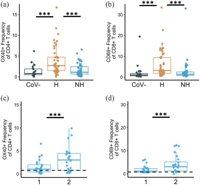
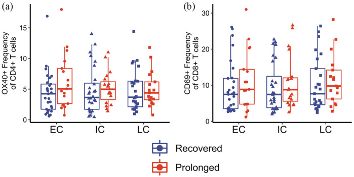
In late 2019, severe acute respiratory syndrome coronavirus 2 (SARS-CoV-2) triggered the global coronavirus disease 2019 (COVID-19) pandemic. Although most infections cause a self-limited syndrome comparable to other upper respiratory viral pathogens, a portion of individuals develop severe illness leading to substantial morbidity and mortality. Furthermore, an estimated 10%-20% of SARS-CoV-2 infections are followed by post-acute sequelae of COVID-19 (PASC), or long COVID. Long COVID is associated with a wide variety of clinical manifestations including cardiopulmonary complications, persistent fatigue, and neurocognitive dysfunction. Severe acute COVID-19 is associated with hyperactivation and increased inflammation, which may be an underlying cause of long COVID in a subset of individuals. However, the immunologic mechanisms driving long COVID development are still under investigation. Early in the pandemic, our group and others observed immune dysregulation persisted into convalescence after acute COVID-19. We subsequently observed persistent immune dysregulation in a cohort of individuals experiencing long COVID. We demonstrated increased SARS-CoV-2-specific CD4+ and CD8+ T-cell responses and antibody affinity in patients experiencing long COVID symptoms. These data suggest a portion of long COVID symptoms may be due to chronic immune activation and the presence of persistent SARS-CoV-2 antigen. This review summarizes the COVID-19 literature to date detailing acute COVID-19 and convalescence and how these observations relate to the development of long COVID. In addition, we discuss recent findings in support of persistent antigen and the evidence that this phenomenon contributes to local and systemic inflammation and the heterogeneous nature of clinical manifestations seen in long COVID.

Keywords: COVID-19; inflammation.


Conflict of interest statement

The author(s) declared the following potential conflicts of interest with respect to the research, authorship, and/or publication of this article: Author NE—intellectual property (IP), “Human neutralizing antibodies against SARS-CoV-2/COVID-19” licensed to the Plantform Corp.

Figures

Figure 1.
Figure 1.
Immune dysregulation of non-hospitalized vs hospitalized individuals during acute SARS-CoV-2 infection. (Blue) Mild illness is associated with a timely immune response, indicated by appropriate activation of the adaptive immune response. This is evident by an increase in cytokine production and upregulation of activation markers on the surface of T cells (non-hospitalized visit 1). While mild illness can lead to a return to baseline function (blue dashed line), we observed that non-hospitalized patients had increased expression of activation and exhaustion markers 1-month post-infection (non-hospitalized visit 2, blue dotted line), indicating persistent immune dysregulation. (Orange/Red) Delayed viral clearance contributes to hyperactivation and worsening of symptoms, leading to hospitalization and mortality. This hyperactivation is associated with upregulation of several activation and exhaustion markers on the surface of T cells, as well as an increase in inflammatory cytokine production (hospitalized/mortality). Furthermore, persistent hyperactivation can lead to eventual exhaustion (hypoactivation) and either recovery (orange dashed line) or mortality (red dashed line). SARS-CoV-2, severe acute respiratory syndrome coronavirus 2.
Figure 2.
Figure 2.
Expression of activation markers on CD4+ and CD8+ T cells during acute COVID-19 infection. (a) OX40 expression on CD4+ T cells and (b) CD69 expression on CD8+ T cells from healthy, pre-pandemic controls (“CoV-”), hospitalized COVID-19-infected samples (“H”), and non-hospitalized COVID-19-infected samples (“NH”). All samples collected during acute infection and early convalescent time points. (c) OX40 expression on CD4+ T cells and (d) CD69 expression on CD8+ T cells from longitudinal non-hospitalized patient samples. Visit number is shown on the x-axis and is represented by “1” and “2” for “Visit 1” and “Visit 2,” respectively. All data modified from Files et al. ***p < 0.001; significance determined by unpaired Wilcoxon tests (a and b) or paired Wilcoxon tests (c and d). COVID-19, corona virus 2019.
Figure 3.
Figure 3.
Expression of activation markers on CD4+ and CD8+ T cells during long COVID. (a) OX40 expression on CD4+ T cells and (b) CD69 expression on CD8+ T cells from patients with prolonged symptoms (long COVID; red) and recovered COVID-19 samples (dark blue). Samples collected at early convalescent (“EC”), intermediate convalescent (“IC”), or late convalescent (“LC”) timepoints. All data modified from Files et al. ***p < 0.001; significance determined by unpaired Wilcoxon tests.
Figure 4.
Figure 4.
Antigen-specific CD4+ T-cell responses are sustained into late convalescence in long COVID-19 patients. (a) OX40+PDL1+ expression on CD4+ T cells following stimulation with S-protein peptide pool from patients with prolonged symptoms (long COVID; red) and recovered COVID-19 samples (dark blue). Samples collected at early convalescent (“EC”), intermediate convalescent (“IC”), or late convalescent (“LC”) timepoints. (b) OX40+PDL1+ expression on CD4+ T cells following stimulation with S-protein peptide pool shown as days post-symptom onset. All data modified from Files et al. **p < 0.01 and *p < 0.05; significance determined by unpaired Wilcoxon tests (a) and by linear mixed effects model (b).
Figure 5.
Figure 5.
Proposed mechanism for how viral reservoirs and dysregulated immune responses contribute to the development of long COVID. Persistent antigen contributes to cell population changes such as a decrease in naïve T and B cells and an increase in effector cells, as well as increased expression of activation and exhaustion markers as the immune system attempts to regulate this chronic activation state. We propose that viral reservoirs contribute to this dysregulation and lead to both localized and systemic inflammation depending on where antigen is located. Several groups have demonstrated antigen persistence in a variety of tissues (e.g., brain, lungs, heart, adipose, gastrointestinal tract) in SARS-CoV-2-infected patients both acutely and chronically. In addition, several groups have demonstrated tissue-specific immune responses and inflammation in relation to corresponding organ-specific symptoms. COVID, coronavirus disease; SARS-CoV-2, severe acute respiratory syndrome coronavirus 2.

Similar articles

Cited by

References

    1. Dong E, Du H, Gardner L.An interactive web-based dashboard to track COVID-19 in real time. Lancet Infect Dis 2020; 20: 533–534. - PMC - PubMed
    1. Ahrenfeldt LJ, Nielsen CR, Möller S, et al.. Burden and prevalence of risk factors for severe COVID-19 disease in the ageing European population–A SHARE-based analysis. Z Gesundh Wiss 2022; 30: 2081–2090. - PMC - PubMed
    1. Anesi GL, Halpern SD, Delgado MK.Covid-19 related hospital admissions in the United States: needs and outcomes. BMJ 2020; 369: m2082. - PubMed
    1. Bohn MK, Hall A, Sepiashvili L, et al.. Pathophysiology of COVID-19: mechanisms underlying disease severity and progression. Physiology (Bethesda) 2020; 35: 288–301. - PMC - PubMed
    1. Chen N, Zhou M, Dong X, et al.. Epidemiological and clinical characteristics of 99 cases of 2019 novel coronavirus pneumonia in Wuhan, China: a descriptive study. Lancet 2020; 395: 507–513. - PMC - PubMed

Publication types