. 2023 Feb 11:28:355-365.
doi: 10.1016/j.omtm.2023.02.003. eCollection 2023 Mar 9.

Specific reprogramming of alpha cells to insulin-producing cells by short glucagon promoter-driven Pdx1 and MafA

Affiliations

Specific reprogramming of alpha cells to insulin-producing cells by short glucagon promoter-driven Pdx1 and MafA

Ping Guo et al. Mol Ther Methods Clin Dev. .

Abstract

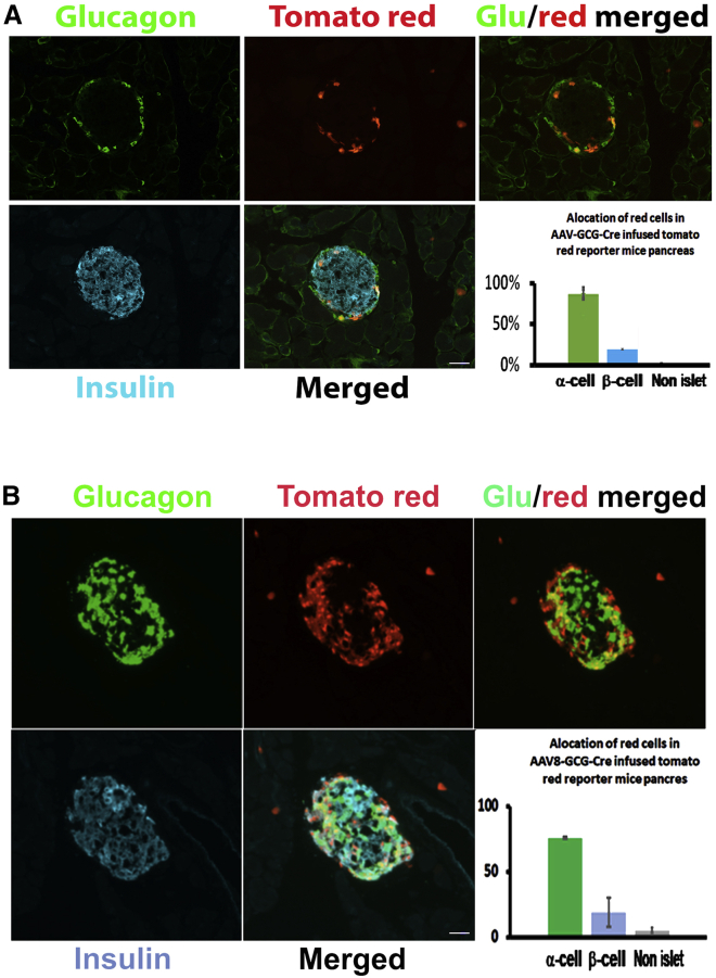
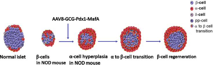
Endogenous reprogramming of pancreas-derived non-beta cells into insulin-producing cells is a promising approach to treat type 1 diabetes (T1D). One strategy that has yet to be explored is the specific delivery of insulin-producing essential genes, Pdx1 and MafA, to pancreatic alpha cells to reprogram the cells into insulin-producing cells in an adult pancreas. In this study, we used an alpha cell-specific glucagon (GCG) promoter to drive Pdx1 and MafA transcription factors to reprogram alpha cells to insulin-producing cells in chemically induced and autoimmune diabetic mice. Our results showed that a combination of a short glucagon-specific promoter with AAV serotype 8 (AAV8) can be used to successfully deliver Pdx1 and MafA to pancreatic alpha cells in the mouse pancreas. Pdx1 and MafA expression specifically in alpha cells were also able to correct hyperglycemia in both induced and autoimmune diabetic mice. With this technology, targeted gene specificity and reprogramming were accomplished with an alpha-specific promotor combined with an AAV-specific serotype and provide an initial basis to develop a novel therapy for the treatment of T1D.

Keywords: adeno-associated virus (AAV) vector; glucagon (GCG) promoter; pancreatic and duodenal homeobox 1 (Pdx1); protein a (MafA); type 1 diabetes (T1D); v-maf musculoaponeurotic fibrosarcoma oncogene family.


Conflict of interest statement

The authors declare no competing interests.

Figures

None
Graphical abstract
Figure 1
Figure 1
T1D mouse alpha cell number increased following disease advancement This figure demonstrates the few remaining beta cells and increased number of alpha cells occupying the islet center. More alpha cells can be observed in the NOD mouse islets as blood glucose levels increased. (A) Normal islet of C57BL/6J (WT). (B) Islet of NOD/ShiLtJ mouse at normal glycemia (NG). (C) Alpha cells tend to be centered in the islet at early onset of diabetes (250 mg/dL). (D and E) Alpha cell distribution in the islets during disease development. (F) Alpha cells occupying the center area of the islet with blood glucose levels at 580 mg/dL. (H–J) Greater proliferation of alpha cells at blood glucose levels of 300 mg/dL (hyperglycemia) of NOD/ShiLtJ mice compared with WT mice. Much more non-glucagon-positive proliferation cells at blood glucose levels of 600 mg/dL compared with early onset of diabetes (J). (K) Quantification of Ki67-positive alpha cells in NOD/ShiLtJ mice islets. ∗p < 0.05. Immunofluorescence staining of Ki 67, glucagon (Glu), and DAPI, ×200, scale bar, 25 μM.
Figure 2
Figure 2
Construction for AAV vector of GCG promoter drive gene A schematic showing the construction of the AAV vector and GCG promoter driving GFP and Cre. For AAV-GCG-Pdx1-MafA, the GCG promoter drives Pdx1 and MafA open reading frames (ORFs), and the GFP tag was connected to the 2A peptide sequence, pAAV-GCG-Pdx1-P2A-MafA-T2A-GFP (A). The Pdx1 and MafA expression of plasmids was determined by western blot. Lanes 1, 2, and 3 are three individual vector-transduced samples (C) and GFP cell transduction efficiency in alpha-TC1 clone 9 cells (B). Min6, Min-6 cell line, a positive control. Bright field and fluorescence images, ×100.
Figure 3
Figure 3
AAV8 GCG promoter drives Pdx-MafA-GFP-specific targeting to alpha cells Specific alpha cell expression of GFP in vivo (A) indicated that Pdx1 and MafA expression was specific to alpha cells compared with AAV8-CMV-GFP virus-infused pancreas (10 days after viral infusion). Immunofluorescence staining, ×200, scale bar, 25 μM. (B) Quantification of GFP-positive cells (more than 96%, p<0.05). RT-PCR images of Pdx1 and MafA after FACS sorting of GFP-positive cells from AAV8-GCG-PM viral-infused islets 2 weeks post viral infusion (C). (D) Western blot of Pdx1 and MafA and ZsGreen isolated from AAV8-GCG-PM viral-infused islets with GFP-positive cells. Lane ms1, ms2 are from mouse 1 and mouse 2; C: AAV-GCG-GFP transduced alpha TC cells as negative control.
Figure 4
Figure 4
AAV8 GCG promoter drives Cre-specific targeting alpha cells in the pancreas of tomato reporter mice The specific alpha cell expression of tomato red in the nuclei of alpha cells by Cre cleaved stop codons in the tomato reporter mouse pancreas via AAV8-GCG-Cre virus infusion. More than 94% alpha cells contain tomato red nuclei, while few beta cells contain tomato red nuclei (p<0.05). These glucagon-positive cells are not beta cells as confirmed by confocal microscope scan and the cleaving of Cre as displayed by red fluorescence. Few non-nuclear red fluorescent cells that were not glucagon positive were found outside of the islets (A). Alpha cells transduced with AAV8-GCG-Cre virus proliferated, and some insulin-positive cells were red and glucagon positive (p<0.05), indicating that the cells transdifferentiated to insulin-producing cells 10 days post viral infusion in the ALX-induced diabetic pancreas (B). Immunofluorescence staining, ×200, scale bar, 25 μM. These results are confirmed by confocal microscope scan. Some cells are insulin and glucagon double-positive (Figure S1).
Figure 5
Figure 5
AAV8 GCG-driven Pdx1 and MafA normalize blood glucose in ALX-induced diabetic mice Blood glucose levels were normalized 3 weeks post AAV8-GCG-PM viral infusion (A). IPGTT showed that glucose tolerance was better than that of the pancreas infused with the AAV-GCG-GFP virus (B). Compared with AAV8-GCG-GFP viral-infused islets (C, e–h) in ALX-induced diabetic mice, ALX islets were normalized by reprogrammed alpha cells into insulin-producing cells in ALX-induced C57/6 mice pancreas (C, i–l). Beta-like cell mass significantly increased compared with the cell mass of AAV8-GCG-GFP viral-infused pancreas (p < 0.01) and up to 65% of WT mice 4 weeks after AAV8-GCG-PM viral infusion (D). Immunofluorescence staining, ×200, scale bar, 25 μM.
Figure 6
Figure 6
AA8 GCG-driven Pdx1 and MafA normalize blood glucose in NOD/ShiLtJ mice with autoimmune diabetes NOD/ShiLtJ mouse blood glucose levels 4 weeks after AAV8-GCG-PM viral infusion (A). Immunohistochemistry of glucagon and insulin in pancreatic tissue of AAV8-GCG-GFP viral-infused NOD/ShiLtJ mice (B, a–d). Pancreatic islets were normalized by reprogramming alpha cells into insulin-producing cells in AAV8-GCG-PM viral-infused NOD/ShiLtJ mice (B, e to l, shows two pancreatic tissue samples). Immunofluorescence staining, ×200, scale bar, 25 μM. IPGTT showed that glucose tolerance was better than that of the pancreas infused with AAV-GCG-GFP (C). Beta-like cell mass was increased at 4 weeks (∗p < 0.01) (D) and maintained until the end of the 30-week experiment. None of the AAV8-GCG-GFP-infused NOD/ShiLtJ mice survived after 6 weeks from viral infusion (D). Concentrations of triglycerides, free fatty acids, and β-hydroxybutyrate in the serum of AAV8-GCG-PM virus-infused mice had normal insulinemia (E). HG, hyperglycemic NOD/ShiLtJ mice; NG, NOD/ShiLtJ normoglycemic; NOD + GCG-PM, AAV8-GCG-PM-infused NOD/ShiLtJ mice. ∗p < 0.01, #p < 0.05.
Figure 7
Figure 7
The GCG promoter drives Pdx1 and MafA in reprogrammed insulin-producing, beta-like cells and reduces immune cell homing to the islets of the pancreas in NOD/ShiLtJ mice CD45-positive immune cells infiltrated the islets of NOD/ShiLtJ mice, but did not infiltrate the AAV8-GCG-PM viral-infused pancreatic islets of NOD/ShiLtJ mice. Immunofluorescence staining, ×200, scale bar, 25 μM.
Figure 8
Figure 8
Schematic of alpha cell reprogramming experiment by GCG promoter combined with AAV viral infusion in ALX-induced and NOD/ShiLtJ diabetic mice

References

    1. Gerrish K., Gannon M., Shih D., Henderson E., Stoffel M., Wright C.V., Stein R. Pancreatic beta cell-specific transcription of the pdx-1 gene. The role of conserved upstream control regions and their hepatic nuclear factor 3beta sites. J. Biol. Chem. 2000;275:3485–3492. doi: 10.1074/jbc.275.5.3485. - DOI - PubMed
    1. Matsuoka T.A., Zhao L., Artner I., Jarrett H.W., Friedman D., Means A., Stein R. Members of the large Maf transcription family regulate insulin gene transcription in islet beta cells. Mol. Cell Biol. 2003;23:6049–6062. doi: 10.1128/MCB.23.17.6049-6062.2003. - DOI - PMC - PubMed
    1. Zhu Y., Liu Q., Zhou Z., Ikeda Y. PDX1, Neurogenin-3, and MAFA: critical transcription regulators for beta cell development and regeneration. Stem Cell Res. Ther. 2017;8:240. doi: 10.1186/s13287-017-0694-z. - DOI - PMC - PubMed
    1. Shapiro A.M.J., Pokrywczynska M., Ricordi C. Clinical pancreatic islet transplantation. Nat. Rev. Endocrinol. 2017;13:268–277. doi: 10.1038/nrendo.2016.178. - DOI - PubMed
    1. Soria B., Gauthier B.R., Martín F., Tejedo J.R., Bedoya F.J., Rojas A., Hmadcha A. Using stem cells to produce insulin. Expet Opin. Biol. Ther. 2015;15:1469–1489. doi: 10.1517/14712598.2015.1066330. - DOI - PubMed