. 2023 Jan 18;12(2):e273-e278.
doi: 10.1016/j.eats.2022.11.002. eCollection 2023 Feb.

Effectivity of the Outside-In Pie-Crusting Technique and an All-Inside Meniscal Repair Device in the Repair of Ramp Lesions

Affiliations

Effectivity of the Outside-In Pie-Crusting Technique and an All-Inside Meniscal Repair Device in the Repair of Ramp Lesions

Koki Kawada et al. Arthrosc Tech. .

Abstract

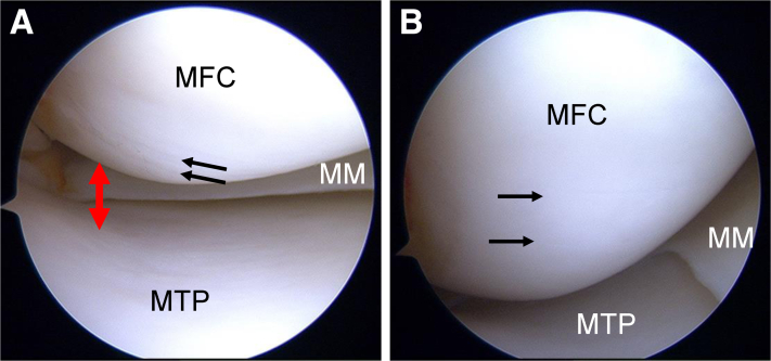
Ramp lesions are characteristic medial meniscus injuries seen in anterior cruciate ligament-injured knees. Anterior cruciate ligament injuries combined with ramp lesions increase the amount of anterior tibial translation and tibial external rotation. Therefore, the diagnosis and treatment of ramp lesions have received increasing attention. However, ramp lesions can be difficult to diagnose on preoperative magnetic resonance imaging. Additionally, ramp lesions are difficult to observe and treat intraoperatively in the posteromedial compartment. Although good results have been reported with the use of a suture hook through the posteromedial portal in the treatment of ramp lesions, the complexity and difficulty of the technique are further problems. The outside-in pie-crusting technique is a simple procedure that can enlarge the medial compartment and facilitate the observation and repair of ramp lesions. After this technique, ramp lesions can be properly sutured, using an all-inside meniscal repair device, without damaging the surrounding cartilage. A combination of the outside-in pie-crusting technique and an all-inside meniscal repair device (with only anterior portals) is effective in the repair of ramp lesions. This Technical Note aims to report in detail the flow of a series of techniques, including our diagnostic and therapeutic methods.


Figures

Fig 1
Fig 1
Preoperative magnetic resonance imaging (MRI) of ramp lesion in right knee. (A) Suspected ramp lesion (arrow) on sagittal view (T2-weighted Dixon sequence). (B) Coronal view (T2-weighted BLADE MRI with fat saturation) showing ramp lesion (arrows).
Fig 2
Fig 2
Arthroscopic view of crevice sign on medial femoral condyle (MFC) and medial compartment before outside-in pie-crusting technique in right knee (supine position, anterolateral portal). This sign indicates instability of the medial meniscus (MM) in anterior cruciate ligament–deficient knees. When this sign is present, MM injuries (especially ramp lesions) are possibly present and require treatment. (A) Small fissures (black arrows) on the MFC are suspected. The view of the posteromedial compartment is extremely narrow (red arrow). (B) Two longitudinal cartilage fissures (arrows) are evident as the knee joint is placed in mild flexion. This is called the crevice sign. (MTP, medial tibial plateau.)
Fig 3
Fig 3
Scope path to intercondylar notch view in right knee (supine position, anterolateral portal). The intercondylar notch view is obtained by passing the scope from the anterolateral portal through to the medial margin of the posterior cruciate ligament (PCL) and the lateral wall of the medial femoral condyle (MFC).
Fig 4
Fig 4
Arthroscopic view of ramp lesion through intercondylar notch view in right knee (supine position, anterolateral portal). (A) Before the outside-in pie-crusting technique is performed, the ramp lesion (black dots) must be confirmed; however, the field that can be observed is narrow, and no space for repair is present. (B) After the outside-in pie-crusting technique is performed, the medial compartment becomes enlarged and the ramp lesion (black dots) can be observed from above. This technique further provides ample space for probing and manipulating all-inside meniscus repair devices. (MFC, medial femoral condyle; MM, medial meniscus.)
Fig 5
Fig 5
Outside-in pie-crusting technique in right knee. The outside-in pie-crusting technique is performed using a standard 18-gauge (1.2 × 40–mm) hypodermic needle. To avoid damage to the meniscus, the needle is targeted between the meniscus and tibial plateau. While the direction of the needle is changed from anterior to posterior, adequate release of the posterior one-third of the medial collateral ligament (MCL) and the posterior oblique ligament (POL) is performed (red arrow). (MM, medial meniscus.)
Fig 6
Fig 6
Arthroscopic view of medial compartment after outside-in pie-crusting technique in right knee (supine position, anterolateral portal). The view of the posteromedial compartment (arrow) is considerably enlarged after the outside-in pie-crusting technique. (MFC, medial femoral condyle; MM, medial meniscus; MTP, medial tibial plateau.)
Fig 7
Fig 7
Rasping of lamp lesion in right knee (supine position, anterolateral portal). The ramp lesion should be rasped before suturing. (MFC, medial femoral condyle; MM, medial meniscus.)
Fig 8
Fig 8
Suturing of ramp lesion using all-inside meniscal repair device (e.g. FAST-FIX) in right knee (supine position, anterolateral portal). (A) By use of the FAST-FIX device, the first anchor is inserted into the meniscus; after the pie-crusting technique, this device can easily be used without damaging the cartilage. (B) The second anchor is inserted into the posterior capsule across the ramp lesion. (MFC, medial femoral condyle; MM, medial meniscus; MTP, medial tibial plateau.)
Fig 9
Fig 9
Adjustment of suture tension using probe in right knee (supine position, anterolateral portal). To avoid over-tightening of the thread, a probe is used to carefully and gradually tighten the thread to the appropriate tension. (MFC, medial femoral condyle; MM, medial meniscus; MTP, medial tibial plateau.)
Fig 10
Fig 10
Arthroscopic view of suturing with all-inside meniscal repair device in right knee (supine position, anterolateral portal). The ramp lesion is shown after the placement of 2 stitches by use of the all-inside meniscal repair device. (MFC, medial femoral condyle; MM, medial meniscus; MTP, medial tibial plateau.)
Fig 11
Fig 11
Arthroscopic view after reconstruction of anterior cruciate ligament in right knee (supine position, anterolateral portal). Anterior cruciate ligament reconstruction is performed using standard procedures, with anatomic double-bundle reconstruction of the semitendinosus tendon. (LFC, lateral femoral condyle; MFC, medial femoral condyle.)

References

    1. Stephen J.M., Halewood C., Kittl C., Bollen S.R., Williams A., Amis A.A. Posteromedial meniscocapsular lesions increase tibiofemoral joint laxity with anterior cruciate ligament deficiency, and their repair reduces laxity. Am J Sports Med. 2016;44:400–408. - PubMed
    1. Malatray M., Raux S., Peltier A., Pfirrmann C., Seil R., Chotel F. Ramp lesions in ACL deficient knees in children and adolescent population: A high prevalence confirmed in intercondylar and posteromedial exploration. Knee Surg Sports Traumatol Arthrosc. 2018;26:1074–1079. - PubMed
    1. Kim S.H., Park Y.B., Won Y.S. An increased lateral femoral condyle ratio is an important risk factor for a medial meniscus ramp lesion including red-red zone tear. Arthroscopy. 2021;37:3159–3165. - PubMed
    1. Nakase J., Asai K., Yoshimizu R., Kimura M., Tsuchiya H. How to detect meniscal ramp lesions using ultrasound. Arthrosc Tech. 2021;10:e1539–e1542. - PMC - PubMed
    1. Murgier J., Hansom D., Clatworthy M. Knee arthroscopy: The “crevice sign,” a new pathognomonic sign for unstable posterior medial meniscal tear in anterior cruciate ligament-deficient knees. Arthrosc Tech. 2020;9:e263–e265. - PMC - PubMed

LinkOut - more resources