

Direct pulp capping procedures - Evidence and practice
- PMID: 36880059
- PMCID: PMC9985044
- DOI: 10.1016/j.jdsr.2023.02.002
Direct pulp capping procedures - Evidence and practice
Abstract
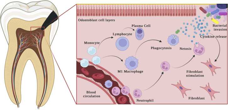
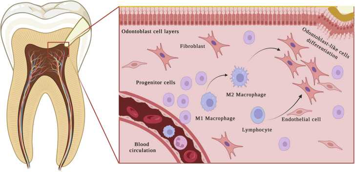
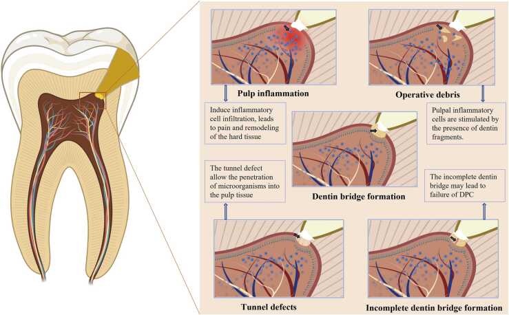
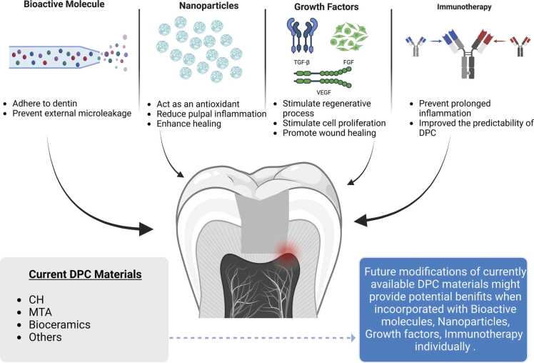
The aim of direct pulp capping (DPC) is to promote pulp healing and mineralized tissue barrier formation by placing a dental biomaterial directly over the exposed pulp. Successful application of this approach avoids the need for further and more extensive treatment. In order to ensure a complete pulp healing with the placement of restorative materials, a mineralized tissue barrier must form to protect the pulp from microbial invasion. The formation of mineralized tissue barrier can only be induced when there is a significant reduction in pulp inflammation and infection. Consequently, promoting the healing of pulp inflammation may provide a favorable therapeutic opportunity to maintain the sustainability of DPC treatment. Mineralized tissue formation was observed as the favorable reaction of exposed pulp tissue against a variety of dental biomaterials utilized for DPC. This observation reveals an intrinsic capacity of pulp tissue for healing. Therefore, this review focuses on the DPC and its healing procedure as well as the materials used for DPC treatment and their mechanisms of action to promote pulpal healing. In addition, the factors that can affect the healing process of DPC, clinical considerations and future perspective has been described.
Keywords: Bioceramics; Calcium hydroxide; Direct pulp capping; Mineral trioxide aggregate; Pulp capping materials; Vital pulp therapy.
© 2023 The Authors.
Conflict of interest statement
None
Figures
References
-
- Cobanoglu N., Alptekin T., Kitagawa H., Blatz M.B., Imazato S., Ozer F. Evaluation of human pulp tissue response following direct pulp capping with a self-etching adhesive system containing MDPB. Dent Mater J. 2021;40:689–696. - PubMed
-
- Komabayashi T., Zhu Q., Eberhart R., Imai Y. Current status of direct pulp-capping materials for permanent teeth. Dent Mater J. 2016;35:1–12. - PubMed
-
- da Rosa W.L., Cocco A.R., Silva T.M.D., Mesquita L.C., Galarca A.D., Silva A.F.D., et al. Current trends and future perspectives of dental pulp capping materials: a systematic review. J Biomed Mater Res B Appl Biomater. 2018;106:1358–1368. - PubMed
-
- Sun H.H., Jin T., Yu Q., Chen F.M. Biological approaches toward dental pulp regeneration by tissue engineering. J Tissue Eng Regen Med. 2011;5:1–16. - PubMed
Publication types
LinkOut - more resources
Full Text Sources
