. 2023 Sep;56(9):e13440.
doi: 10.1111/cpr.13440. Epub 2023 Mar 7.

Piezo1-mediated M2 macrophage mechanotransduction enhances bone formation through secretion and activation of transforming growth factor-β1

Affiliations

Piezo1-mediated M2 macrophage mechanotransduction enhances bone formation through secretion and activation of transforming growth factor-β1

Guanhui Cai et al. Cell Prolif. 2023 Sep.

Abstract

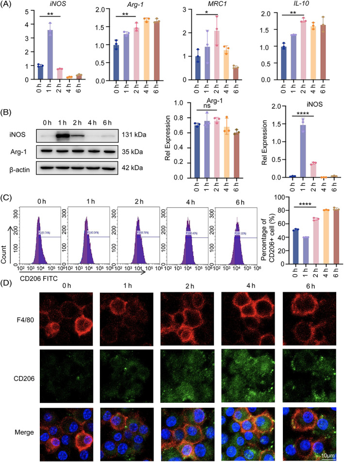
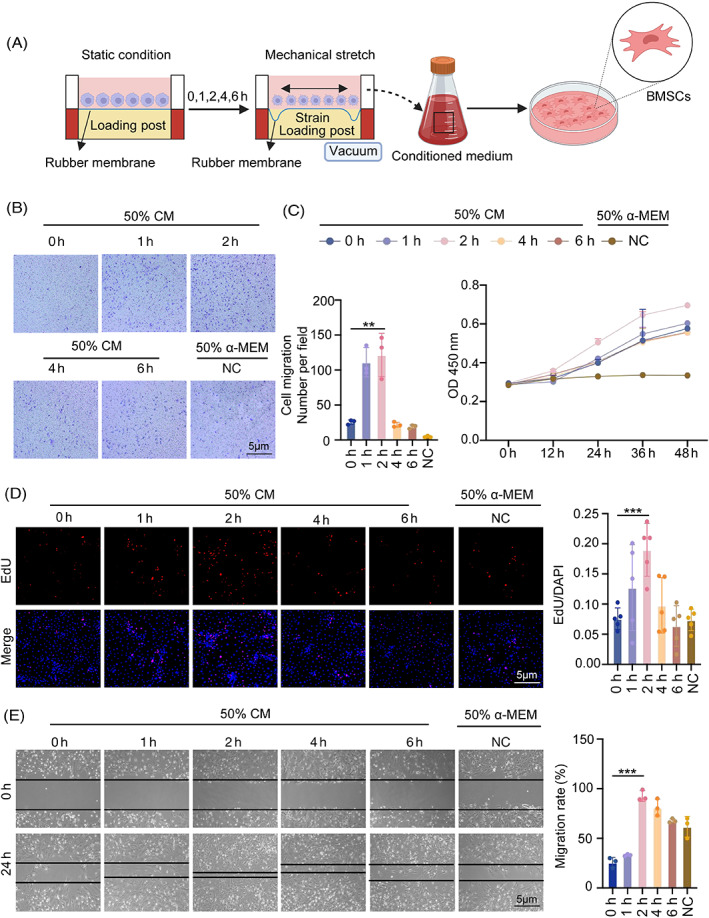
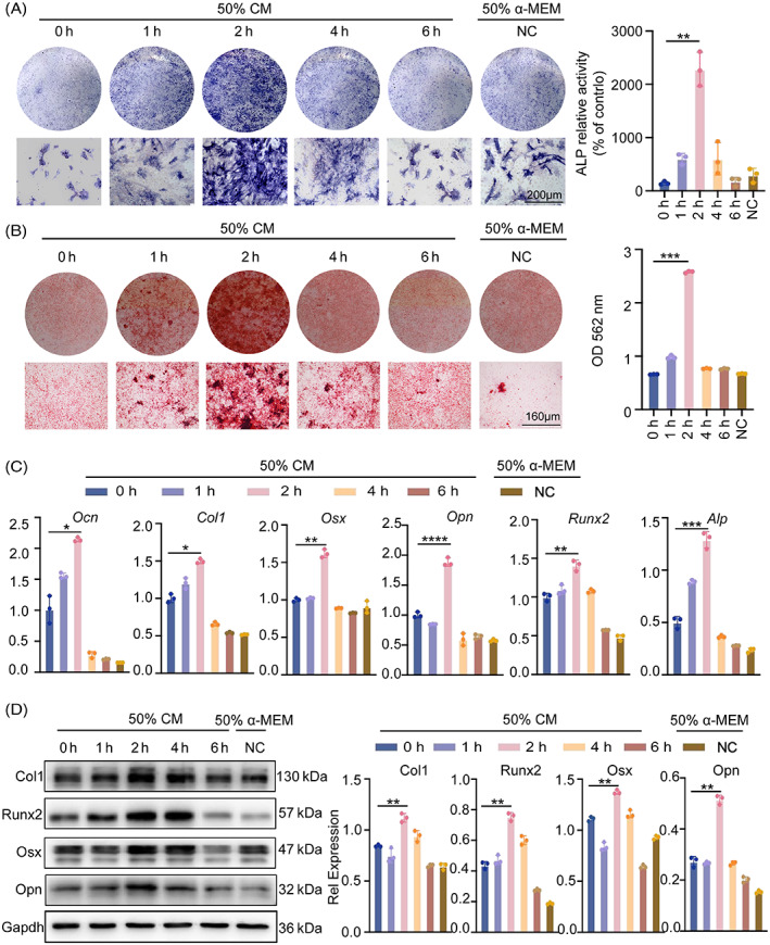
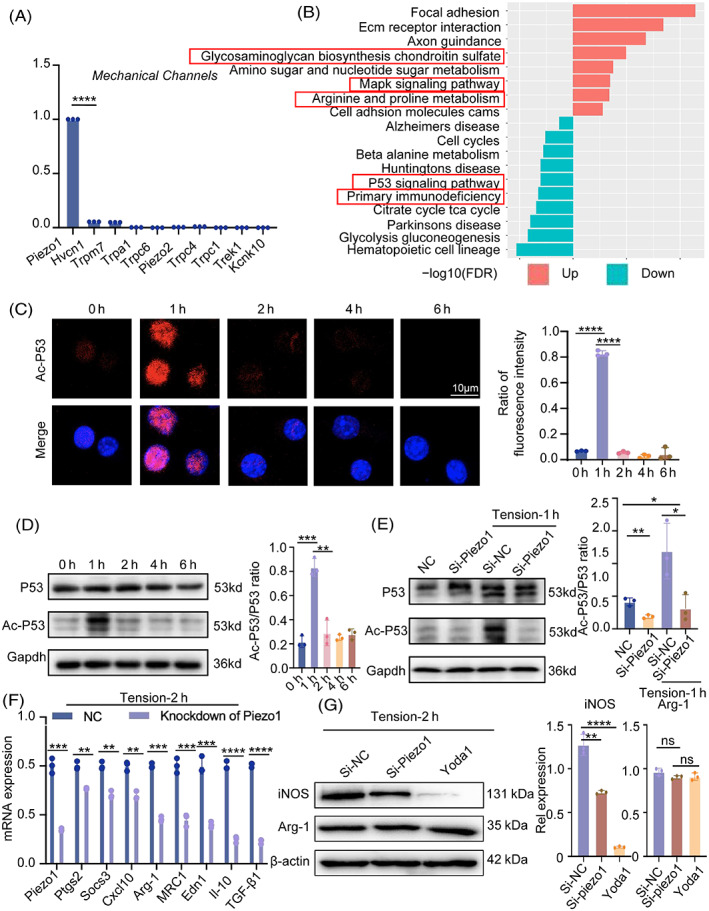
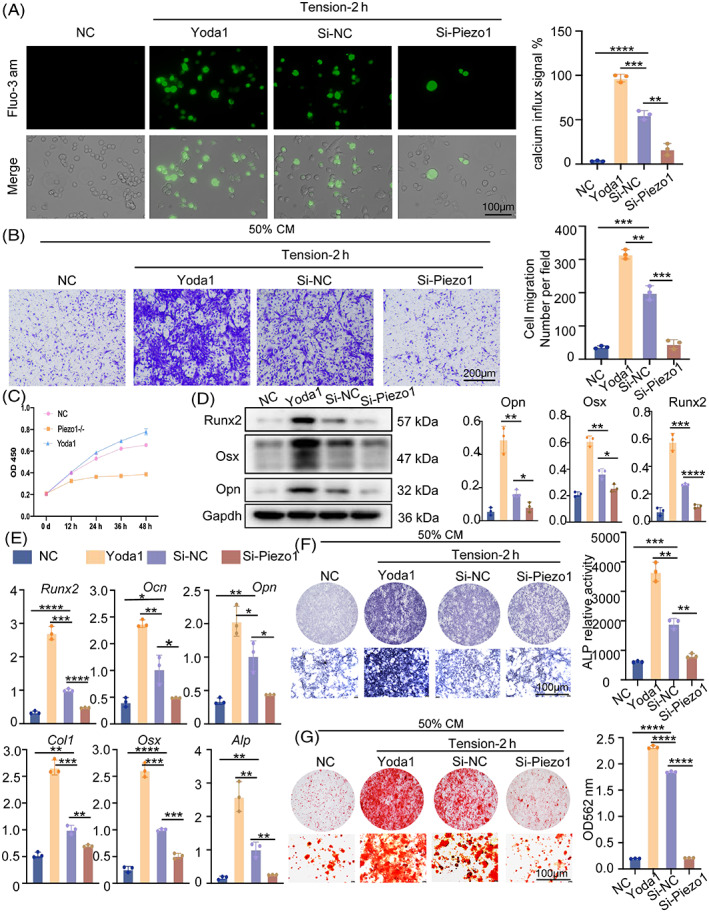
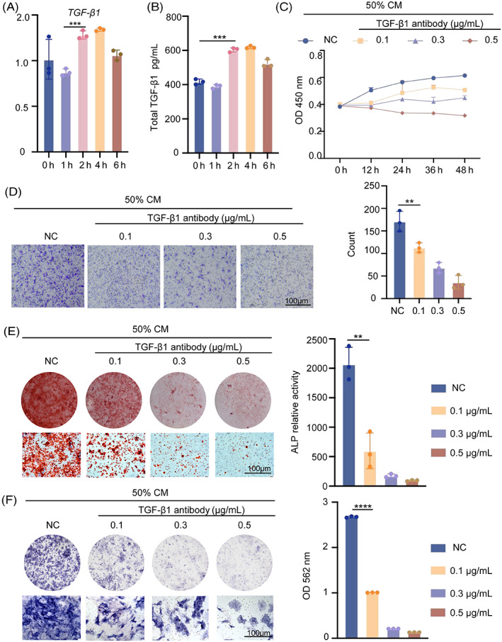
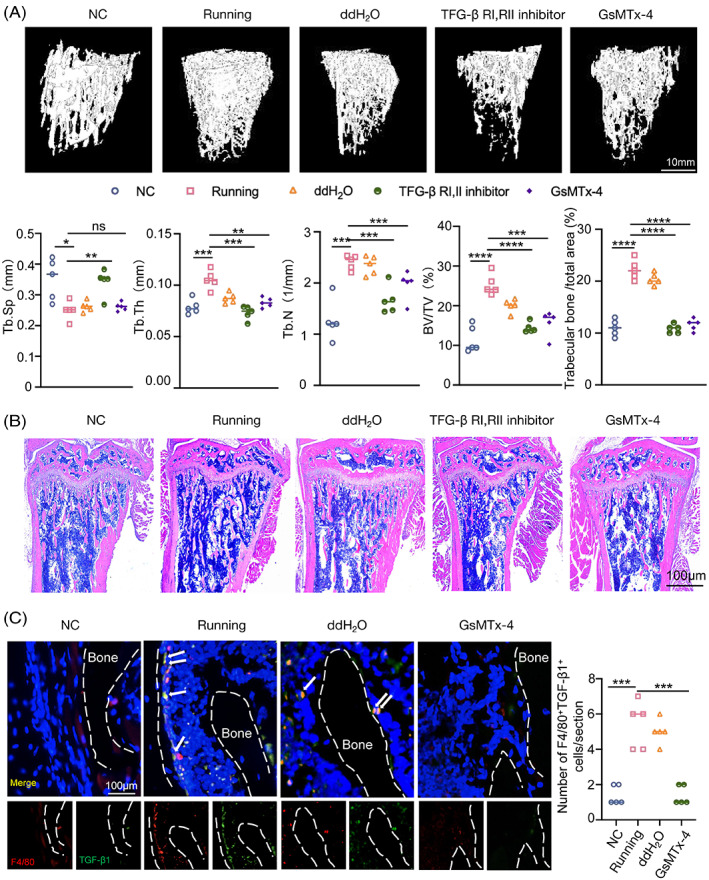
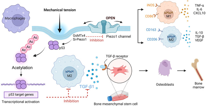
Macrophages are multifunctional immune system cells that are essential for the mechanical stimulation-induced control of metabolism. Piezo1 is a non-selective calcium channel expressed in multifarious tissues to convey mechanical signals. Here, a cellular model of tension was used to study the effect of mechanical stretch on the phenotypic transformation of macrophages and its mechanism. An indirect co-culture system was used to explore the effect of macrophage activation on bone marrow mesenchymal stem cells (BMSCs), and a treadmill running model was used to validate the mechanism in vivo for in vitro studies. p53 was acetylated and deacetylated by macrophages as a result of mechanical strain being detected by Piezo1. This process is able to polarize macrophages towards M2 and secretes transforming growth factor-beta (TGF-β1), which subsequently stimulates BMSCs migration, proliferation and osteogenic differentiation. Knockdown of Piezo1 inhibits the conversion of macrophages to the reparative phenotype, thereby affecting bone remodelling. Blockade of TGF-β I, II receptors and Piezo1 significantly reduced exercise-increased bone mass in mice. In conclusion, we showed that mechanical tension causes calcium influx, p53 deacetylation, macrophage polarization towards M2 and TGF-β1 release through Piezo1. These events support BMSC osteogenesis.


Conflict of interest statement

The authors declare no conflict of interest.

Figures

FIGURE 1
FIGURE 1
Mechanical tension induces macrophage polarization. (A) Real‐time RT‐PCR was used to assess the mRNA levels of iNOS, Arg‐1, MRC1 and IL‐10 in RAW264.7 cells as a result of the 0–6 h of tension. (B) Western blot was used to identify the protein expression of iNOS and Arg‐1 in RAW264.7 cells under stress for 0–6 h. GAPDH was utilized as the standard to quantify iNOS and Arg‐1. (C) The expression of CD206 in RAW264.7 cells under tension was detected by flow cytometry, and the statistical results were displayed. (D) M2 macrophages in RAW264.7 cells under the tension of 0–6 h were visualized using immunofluorescence staining of F4/80 (red) and CD206 (green). The cell nuclei were detected with DAPI. Scale bar: 10 μm. Data represent the mean ± SD. *p < 0.05, **p < 0.01, ***p < 0.001.
FIGURE 2
FIGURE 2
Macrophage‐conditioned medium under tension promotes the proliferation and migration of BMSCs. (A) Diagram of macrophages under tension with the Flexcell® FX‐5000™Tension System. (B) Effects of conditioned medium of macrophages under 0–6 h tension on vertical migration of BMSCs are demonstrated by ×4 micrographs of BMSCs in different groups at the bottom of the migration transwell culture chamber and a histogram of cell counts based on ×4 magnification fields. (C) Cell Counting Kit 8 (CCK8) assays were performed to explore BMSCs proliferation following treatment with the macrophage‐conditioned medium under 0–6 h of tension (magnification ×100). (D) Additionally, BMSCs proliferation was investigated using 5‐ethynyl‐2′‐deoxyuridine (EdU) tests (magnification ×100). (E) Effects of conditioned medium on lateral migration, as shown by the initial and final (24 h) micrographs of the scratch in the wound healing assay. Bar graph of final distance/initial distance rate of each group. Data represent the mean ± SD. *p < 0.05, **p < 0.01, ***p < 0.001.
FIGURE 3
FIGURE 3
Macrophage‐derived medium promotes osteogenic differentiation of BMSC. (A) In osteogenic differentiation medium, BMSCs treated with macrophage supernatants were induced to undergo osteogenesis. ALP expression in BMSCs was measured by the ALP staining method. Gross scanning images are shown at the top (scale bar: 1 mm), expanded images are shown at the bottom (magnification: ×250, scale bar: 160 μm), and a quantification of the gross scanning images is shown on the right. (B) The osteogenic differentiation of BMSCs was evaluated by Alizarin Red staining after 21 days. (C) Ocn, Col1, Osx, Opn, Runx2 and Alp in BMSCs were measured by real‐time RT‐PCR. GAPDH was used for normalization. (D) The protein expression of Col1, Runx2, Osx and Opn in BMSCs with conditioned medium were detected by western blot. GAPDH was used for normalization. Data represent the mean ± SD. *p < 0.05, **p < 0.01, ***p < 0.001.
FIGURE 4
FIGURE 4
Piezo1 is required for the mechanically induced M2 polarization. (A) mRNA levels of calcium channels in RAW264.7 cells were determined by RT‐PCR. (B) Differential signalling pathways were analysed from the database (GSE139121). (C) Immunofluorescent staining of acetyl‐p53 (red) in RAW264.7 cells under the tension of 0–6 h. Blue indicates DAPI staining of nuclei. Scale bar: 10 μm. (D) Western blot was used to identify the p53 and acetyl‐p53 protein expression in RAW264.7 cells as a result of 0–6 h of tension. GAPDH was used for normalization. (E) In the control or Piezo1 knock‐down groups, p53 and acetyl‐p53 protein expression were tested under static or tension conditions. (F) Piezo1, Ptgs2, Socs3, Cxcl10, Arg‐1, MRC1, Edn1, IL‐10 and TGF‐β1 were detected by qPCR in control or Piezo1 knock‐down groups that had been cultivated under tension for 2 h. (G) Western blotting was used to identify the protein expression of iNOS and Arg‐1 in three groups (the control group, the Piezo1‐knockdown group and the Yoda1‐administered group) during 2 h of tension. Data represent the mean ± SD. *p < 0.05, ** p < 0.01, ***p < 0.001.
FIGURE 5
FIGURE 5
Piezo1 knockdown hampers the proliferation, migration and osteogenic differentiation of BMSCs. (A) Fluorescence intensity of Ca2+ after Piezo1 knockdown or treatment with 1 μmol/L Yoda1 as measured by the Fluo‐3 AM probe under a fluorescence microscope. (B) Effects of 1 μmol/L Yoda1 or knockdown of Piezo1 in macrophages on vertical migration of BMSCs under static 2‐h tension, as shown by ×4 micrographs of BMSCs in different groups at the bottom of the migration transwell culture chamber. (C) Cell Counting Kit 8 (CCK8) was performed to explore BMSCs proliferation following treatment with 1 μmol/L Yoda1 or knockdown of Piezo1. (D) The protein expression of Runx2, Osx and Opn in BMSCs were detected by western blot. GAPDH was used for normalization. (E) Col1, Osx, Runx2, Ocn, Opn and Alp in BMSCs were measured by real‐time RT‐PCR. GAPDH was used for normalization. (F) ALP expression in BMSCs was measured by the ALP staining method. The top are gross scanning images (scale bar: 1 mm), and the lower are enlarged images (magnification: ×250, scale bar: 160 μm). (G) After 21 days, the osteogenic differentiation of BMSCs was evaluated by Alizarin Red staining. The top are gross scanning images (scale bar: 1 mm), and the lower are enlarged images (magnification: ×250, scale bar: 160 μm). Data represent the mean ± SD. *p < 0.05, **p < 0.01, ***p < 0.001.
FIGURE 6
FIGURE 6
M2 macrophages promote the osteogenic differentiation of BMSCs by secreting TGF‐β1. (A) TGF‐β1 in RAW264.7 cells was measured by real‐time RT‐PCR. GAPDH was used for normalization. (B) TGF‐β1 secretion from macrophages under various stress situations is measured using an ELISA. (C) Cell Counting Kit 8 (CCK8) was used to investigate BMSCs proliferation after treatment with various concentrations of TGF‐β1 antibody. (D) TGF‐β1 antibody effects on BMSC vertical migration, as indicated by four micrographs of BMSCs in separate groups at the bottom of the migration transwell culture chamber. (E) BMSCs treated with TGF‐β1 antibody were stimulated into osteogenesis in osteogenic differentiation medium. The ALP staining approach was used in BMSCs. The top photos are gross scanning images (scale bar: 160 μm), whereas the bottom images are expanded images (magnification: 250, scale bar: 160 μm). (F) The osteogenic differentiation of BMSCs was assessed by Alizarin Red staining after 21 days. The photos on top are gross scanning images (scale bar: 160 mm), while the images on the bottom are expanded images (magnification: ×250, scale bar: 160 μm). Data represent the mean ± SD. *p < 0.05, **p < 0.01, ***p < 0.001.
FIGURE 7
FIGURE 7
Knockdown of TGF‐β1 and Piezo1 both reduced bone remodelling in running mice. (A) μ‐CT images of the distal tibia trabecular bones isolated from 10‐week‐old male WT‐mice and mice that had been treated with TGF‐β receptor I (RI), receptor II (RII) inhibitor and Piezo1 inhibitor. μ‐CT analysis of interesting region for bone volume per tissue volume (BV/TV), trabecular thickness (Tb.Th), trabecular number (Tb.N), trabecular spacing (Tb.Sp). (B) Representative HE‐staining images of the tibial metaphysis after 1 month of exercise. (C) Representative immunofluorescence staining and quantification of F4/80+ (red) TGF‐β1+ (green) cells on the distal tibia. Scale bar, 100 μm. White arrows indicated F4/80+ and TGF‐β1+ cells.
FIGURE 8
FIGURE 8
Mechanism diagram. Piezo1‐mediated mechanical tension stimulates macrophage polarization and secretion of TGF‐β1, promoting osteogenic differentiation. Acetylation and deacetylation of p53 play a major role in this process.

Similar articles

Cited by

References

    1. Sims NA, Martin TJ. Coupling the activities of bone formation and resorption: a multitude of signals within the basic multicellular unit. Bonekey Rep. 2014;3:481. doi:10.1038/bonekey.2013.215 - DOI - PMC - PubMed
    1. Michalski MN, McCauley LK. Macrophages and skeletal health. Pharmacol Ther. 2017;174:43‐54. doi:10.1016/j.pharmthera.2017.02.017 - DOI - PMC - PubMed
    1. Bleriot C, Chakarov S, Ginhoux F. Determinants of resident tissue macrophage identity and function. Immunity. 2020;52(6):957‐970. doi:10.1016/j.immuni.2020.05.014 - DOI - PubMed
    1. Atcha H, Jairaman A, Holt JR, et al. Mechanically activated ion channel Piezo1 modulates macrophage polarization and stiffness sensing. Nat Commun. 2021;12(1):3256. doi:10.1038/s41467-021-23482-5 - DOI - PMC - PubMed
    1. Adams S, Wuescher LM, Worth R, Yildirim‐Ayan E. Mechano‐immunomodulation: mechanoresponsive changes in macrophage activity and polarization. Ann Biomed Eng. 2019;47(11):2213‐2231. doi:10.1007/s10439-019-02302-4 - DOI - PMC - PubMed