. 2023 Apr 3;222(4):e202201027.
doi: 10.1083/jcb.202201027. Epub 2023 Feb 28.

The p97/VCP segregase is essential for arsenic-induced degradation of PML and PML-RARA

Affiliations

The p97/VCP segregase is essential for arsenic-induced degradation of PML and PML-RARA

Ellis G Jaffray et al. J Cell Biol. .

Abstract

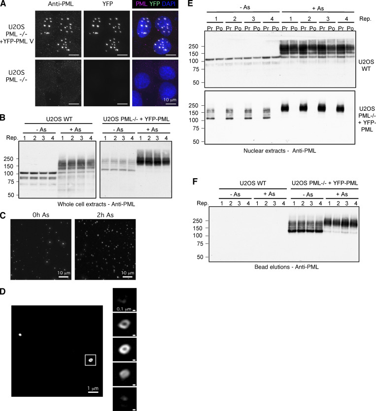
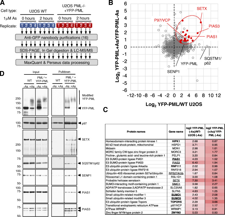
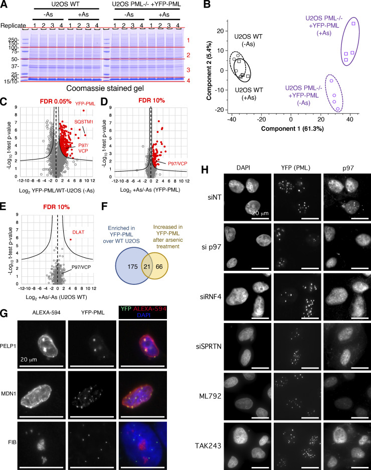
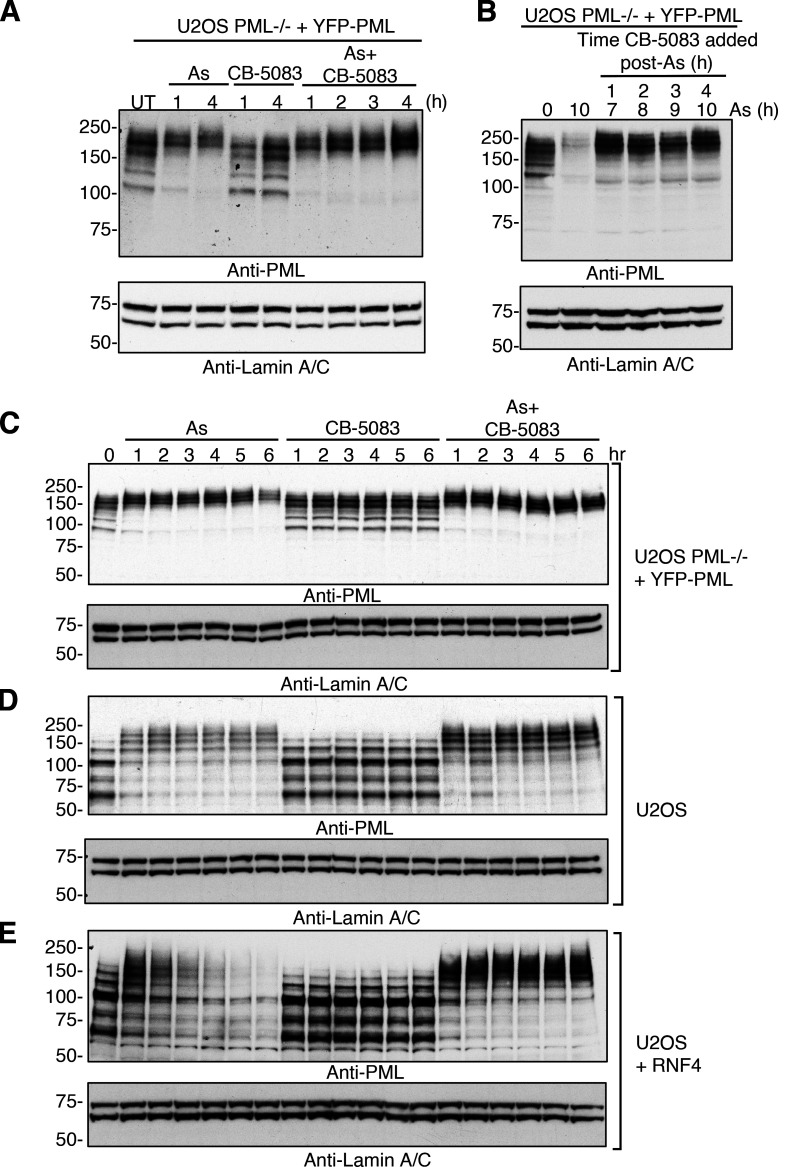
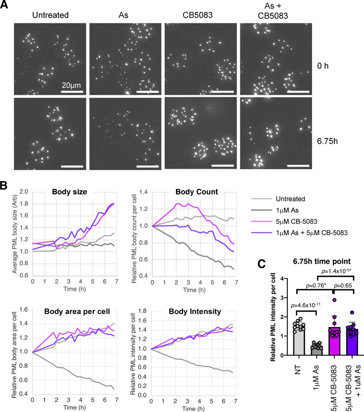
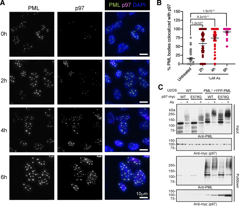
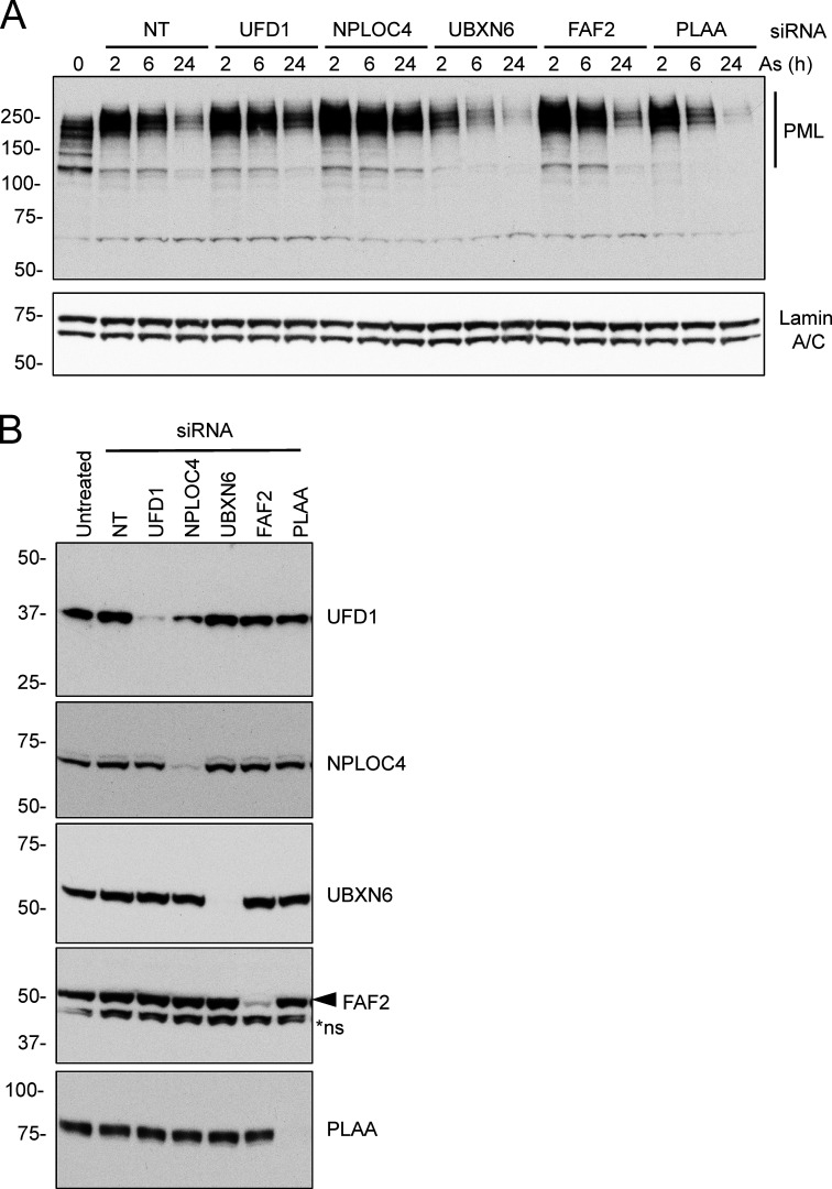
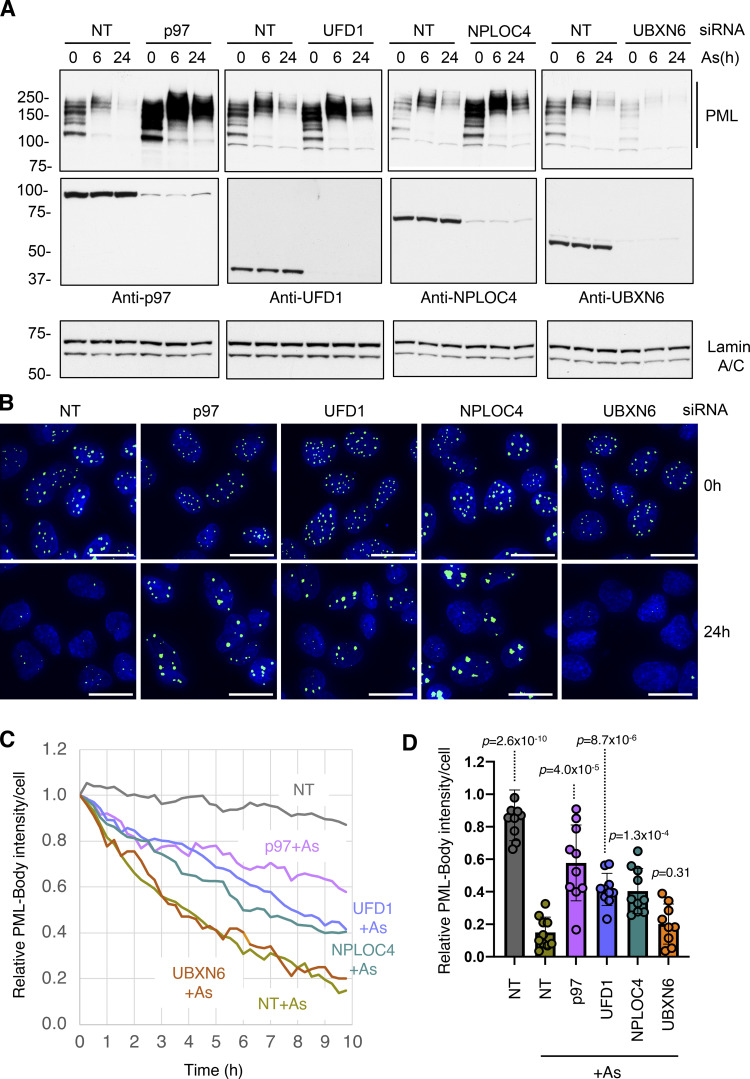
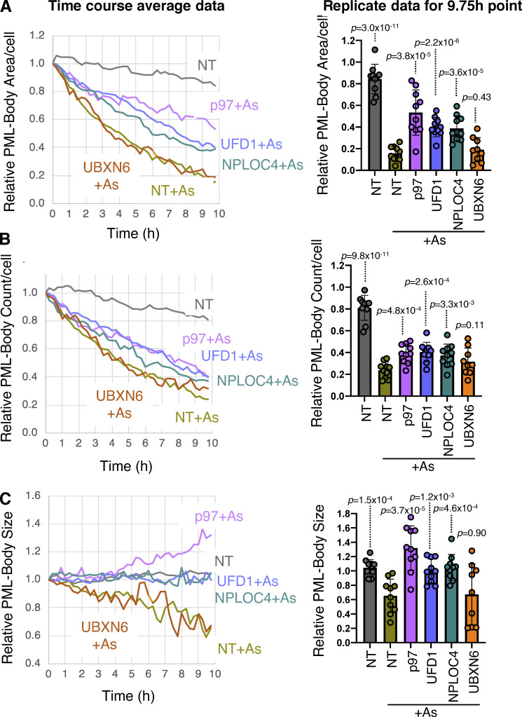
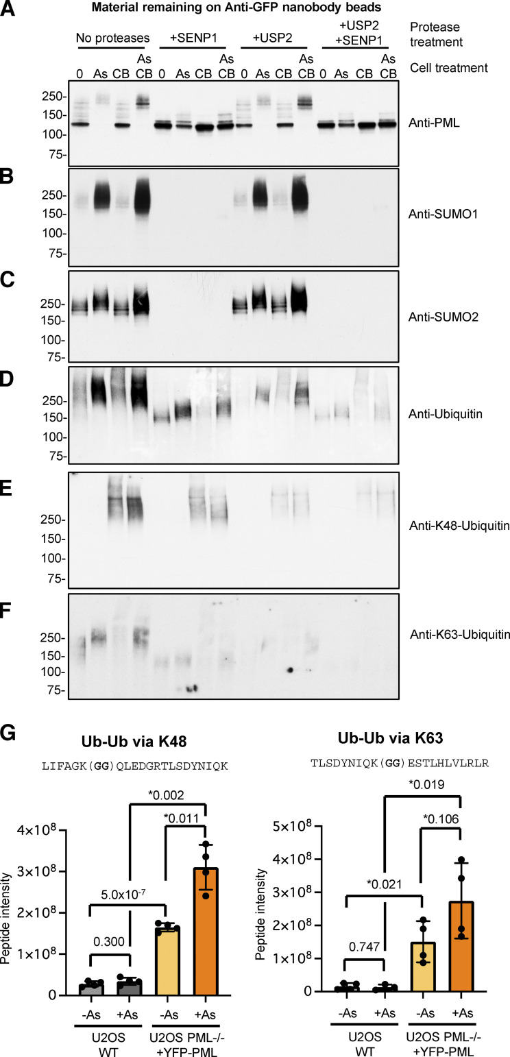
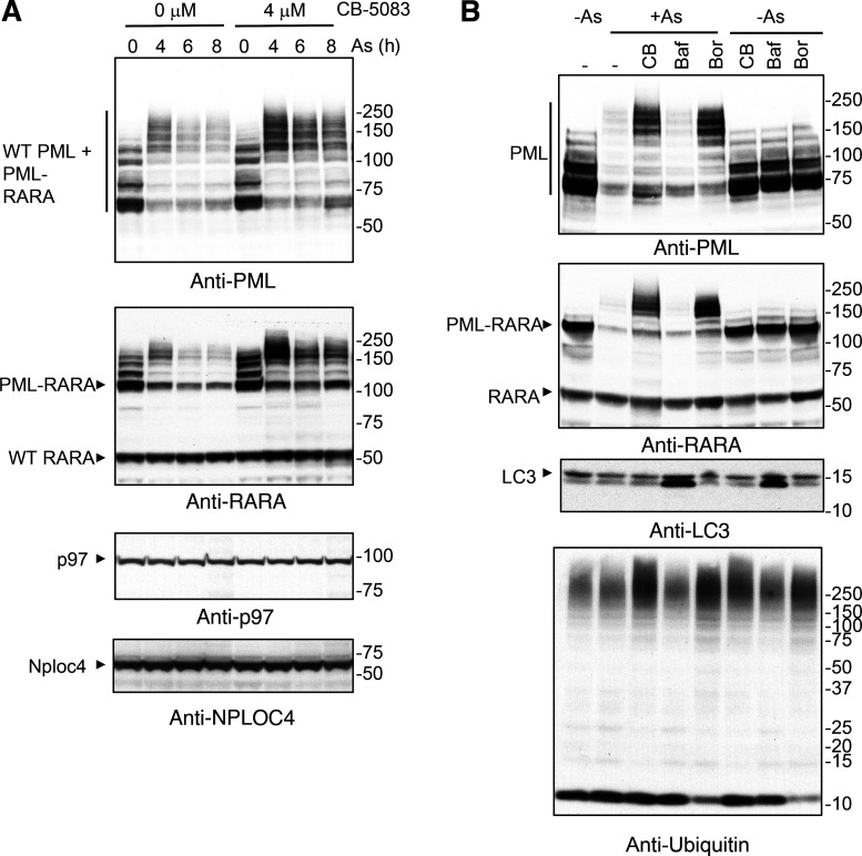
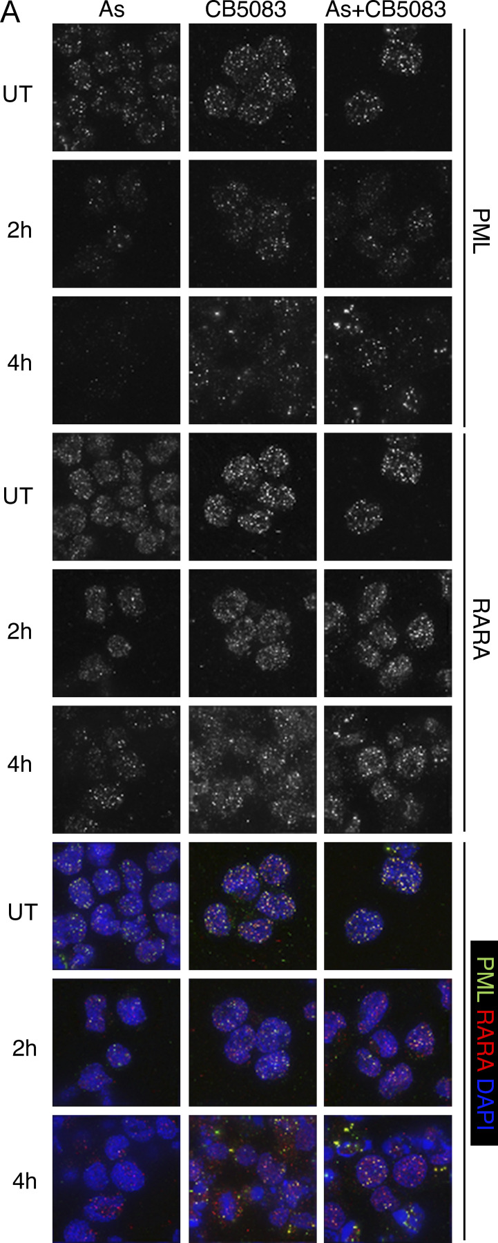
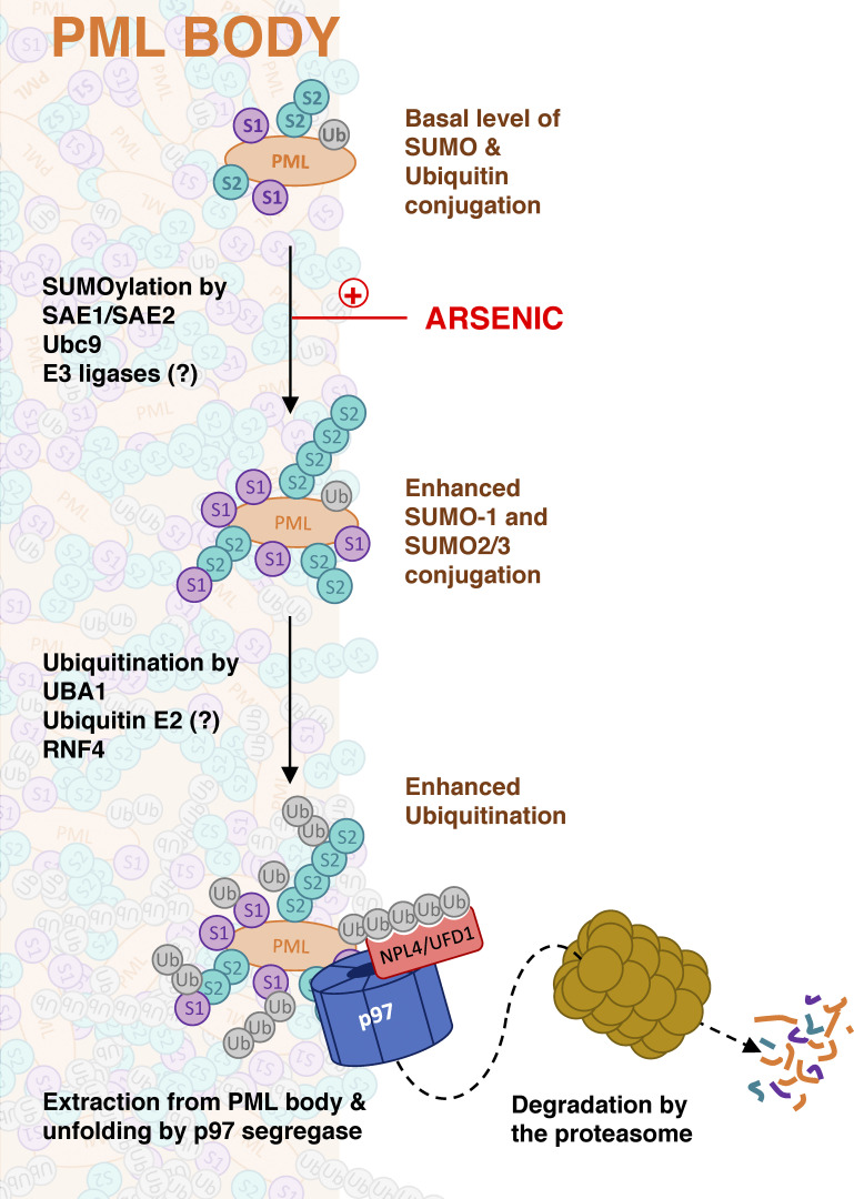
Acute Promyelocytic Leukemia is caused by expression of the oncogenic Promyelocytic Leukemia (PML)-Retinoic Acid Receptor Alpha (RARA) fusion protein. Therapy with arsenic trioxide results in degradation of PML-RARA and PML and cures the disease. Modification of PML and PML-RARA with SUMO and ubiquitin precedes ubiquitin-mediated proteolysis. To identify additional components of this pathway, we performed proteomics on PML bodies. This revealed that association of p97/VCP segregase with PML bodies is increased after arsenic treatment. Pharmacological inhibition of p97 altered the number, morphology, and size of PML bodies, accumulated SUMO and ubiquitin modified PML and blocked arsenic-induced degradation of PML-RARA and PML. p97 localized to PML bodies in response to arsenic, and siRNA-mediated depletion showed that p97 cofactors UFD1 and NPLOC4 were critical for PML degradation. Thus, the UFD1-NPLOC4-p97 segregase complex is required to extract poly-ubiquitinated, poly-SUMOylated PML from PML bodies, prior to degradation by the proteasome.


Conflict of interest statement

Disclosures: The authors declare no competing interests exist.

Figures

Figure S1.
Figure S1.
Analysis of PML bodies purified from cultured cells. (A) Fluorescence microscopy comparing U2OS PML−/− cells (lower) with the U2OS PML−/− + YFP-PML cells (upper). Images captured using chicken anti-PML with Texas Red secondary (left panels) or YFP fluorescence (center panels). Right panels show the two channels merged with DAPI. Scale bars are 10 μm. (B) U2OS WT and U2OS PML−/− + YFP-PML cells in quadruplicate were either untreated (−As) or treated with arsenic for 2 h (+As) and lysates analyzed by Western blotting with an antibody to PML. (C) Fluorescence microscopy of nuclear extracts from U2OS PML−/− + YFP-PML cells either untreated (−As) or treated with arsenic for 2 h (+As). (D) Left panels show a widefield and right panels show four z sections through a single PML body (untreated). (E) Anti-PML Western blots of nuclear extracts either prior (Pr) to or post (Po) incubation with anti-GFP nanobody beads. (F) Anti-PML Western blot of material eluted from anti-GFP nanobody beads. Molecular weight markers are indicated in kD. Source data are available for this figure: SourceData FS1.
Figure 1.
Figure 1.
Proteomic analysis of arsenic-induced PML body associated proteins. (A) Overview of the proteomic experimental design. For each condition, n = 4 separate experiments. (B) Scatter plot of log2 ratios comparing protein abundance in anti-GFP nanobody purifications from YFP-PML expressing PML−/− U2OS cells with those from WT U2OS cells (x-axis) and comparing abundance in purifications form YFP-PML cells during 1 μM arsenic treatment with the same purifications from cells not exposed to arsenic (y-axis). Data for 2,157 proteins shown. Red markers are proteins defined as significantly different in both comparisons (see Fig. S2). (C) Summary data for the 21 proteins marked red in B. Underlined gene names were identified previously as PML proximal proteins (Barroso-Gomila et al., 2021), and bold entries are described in the Human Protein Atlas (Thul et al., 2017; http://proteinatlas.org) as belonging to PML body. (D) Anti-GFP nanobody purifications were prepared from the indicated cells after 6 h 1 μM arsenic (+As) or untreated (−As). Both inputs and bead elutions were Western blotted for the indicated species. Molecular weight markers are indicated in kD. Source data are available for this figure: SourceData F1.
Figure S2.
Figure S2.
VCP/p97 interaction with PML increases during arsenic treatment. (A) Coomassie stained SDS-PAGE gel fractionating anti-GFP affinity purifications from the indicated cell types with or without arsenic treatment (As). Gels were sliced into four sections per lane (as indicated) for in-gel tryptic digestion. Molecular weight markers are indicated in kD. (B) Principal component analysis of intensity data for 2,156 identified proteins. (C–E) Scatter plots showing fold change versus two-tailed student’s t test P value for the comparisons indicated on the x-axes (n = 4). FDR filtering values are indicated above each chart. (F) Venn diagram showing overlap between significant changers for parts C and D. The intersection is highlighted in Fig. 1 B. (G) Immunofluorescence analysis of PELP, MDN1, and FIB in U2OS PML−/− + YFP-PML using Alexa-594 secondary antibodies (left) and YFP fluorescence (center). Merged images with DAPI staining are shown (right panels). (H) In U2OS PML−/− + YFP-PML p97, RNF4 or SPRTN levels were ablated using siRNA or the SUMO and ubiquitin E1 enzymes inhibited with ML792 or TAK243, followed by 6 h treatment with arsenic. YFP-PML colocalization with p97 was measured by fluorescence and immunofluorescence. Source data are available for this figure: SourceData FS2.
Figure 2.
Figure 2.
Pharmacological inhibition of p97 interferes with arsenic-induced degradation of PML. (A) U2OS PML−/− + YFP-PML cells were either untreated (UT) or treated with 1 μM arsenic (As), 5 μM CB-5083, or a combination of arsenic and CB-5083 for the times indicated. Cell lysates were analyzed by Western blotting using an antibody to PML or Lamin A/C. (B) U2OS PML−/− + YFP-PML cells were treated with 1 μM arsenic for the indicated times and 5 μM CB-5083 added at indicated times after the start of arsenic treatment. Cell lysates were analyzed by Western blotting using an antibody to PML or Lamin A/C. (C–E) U2OS PML−/− + YFP-PML cells (C), U2OS cells (D), or U2OS + RNF4 cells (E) were either untreated (UT) or treated with 1 μM arsenic (As), 5 μM CB-5083, or a combination of arsenic and CB-5083 for the times indicated. Cell lysates were analyzed by Western blotting using an antibody to PML or Lamin A/C. Molecular weight markers are indicated in kD. Source data are available for this figure: SourceData F2.
Figure 3.
Figure 3.
Inhibition of p97 alters arsenic-induced changes to PML body number, morphology, and content. U2OS PML−/− + YFP-PML cells were either untreated or treated with 1 μM arsenic (As), 5 μM CB-5083, or a combination of arsenic and CB-5083 and analyzed by time-lapse fluorescent imaging for 6.75 h. 10 videos were collected for each condition (one video from each condition is shown in Videos 1, 2, 3, and 4). (A) Stills from the start (0 h) and the end (6.75 h) of the movies. Scale bars are 20 μm. (B) The average PML body size, number of PML bodies per cell, total area of PML bodies, and total intensity of PML bodies was plotted as a function of time (n = 10 fields of view). (C) Comparison of relative PML intensity per cell after 6.75 h treatment as indicated. Columns represent average and error bars are 1 SD. P values are two-tailed student t tests assuming equal variance or unequal variance (*). Each data point is derived from a single field of view taken from the time course shown in B (n = 10). Source data are available for this figure: SourceData F3.
Figure 4.
Figure 4.
P97 is recruited to PML bodies in response to arsenic. U2OS PML−/− + YFP-PML cells were treated with arsenic for the times indicated, pre-extracted as described in Materials and methods, fixed, and stained with an antibody to p97. (A) Images of p97 (green), YFP-PML (red), and DAPI (blue) are shown. Scale bars are 10 μm. (B) The percentage of PML bodies colocalized with p97 was plotted against time of arsenic treatment. Chart shows individual cell data. Bars represent mean with SEM. P values are two-tailed Mann-Whitney U test derived by comparison with untreated. Cell count n = 41 (untreated), n = 38 (2 h), n = 38 (4 h), and n = 25 (6 h). (C) WT U2OS cells or PML−/− + YFP-PML U2OS cells were transfected with plasmids expressing myc-p97 constructs of either WT or E578Q mutant form and either treated or not with 1 μM arsenic for 6 h. Nuclear extracts (Input) or YFP-PML purifications (Pulldown) were blotted for PML or Myc. Molecular weight markers are indicated in kD. Source data are available for this figure: SourceData F4.
Figure S3.
Figure S3.
Identification of UFD1 and NPLOC4 as the p97 cofactors involved in arsenic-induced PML degradation. U2OS PML−/− + YFP-PML cells were treated with NT or with siRNAs to the indicated p97 cofactors and exposed to arsenic for the times indicated. (A) Cell lysates were analyzed by Western blotting with antibodies to PML and Lamin A/C. (B) Western blotting for the indicated p97 cofactors in whole-cell extracts from cells treated with the indicated siRNAs. Molecular weight markers are indicated in kD. Source data are available for this figure: SourceData FS3.
Figure 5.
Figure 5.
p97 cofactors UFD1 and NPLOC4 are required for arsenic-induced degradation of PML. U2OS PML−/− + YFP-PML cells were treated with NT siRNA or siRNAs to p97 cofactors and exposed to arsenic. (A) Western blot analysis with antibodies to PML, p97 cofactors, or Lamin A/C. Molecular weight markers are indicated in kD. (B) Immunofluorescence analysis of YFP-PML in response to arsenic. Scale bars are 20 μm. (C) Time-lapse fluorescent imaging for 9.75 h after exposure to arsenic. 10 videos were collected for each condition (n = 10; example videos in Videos 5, 6, 7, 8, 9, and 10). Average relative PML body intensity per cell was plotted versus time. (D) Relative PML intensity per cell of cells treated with NT or siRNAs to p97 cofactors after 10 h arsenic (As) treatment. P values are comparisons with NT + As using two-tailed unpaired Student’s t test assuming similar deviations. Each data point is a single field of view taken from C (n = 10). Source data are available for this figure: SourceData F5.
Figure S4.
Figure S4.
UFD1 and NPLOC4 influence the nuclear distribution of PML. U2OS PML−/− + YFP-PML cells were treated with NT siRNA or with siRNAs to the indicated p97 cofactors, exposed to arsenic and analyzed by time-lapse fluorescent imaging for 9.75 h. PML body parameters were plotted as a function of time (left panels; average—n = 10). In the right panels PML body parameters are compared in cells treated with NT siRNA or with siRNAs to the indicated p97 cofactors after 9.75 h arsenic treatment (average value ± SD). P values are two-tailed Student’s t test assuming similar data deviations comparing with NT + As. Each data point is a single field of view taken from the time course (n = 10). (A) Relative PML body area per cell. (B) Relative PML body number per cell. (C) Relative PML body size.
Figure 6.
Figure 6.
P97 inhibition leads to the accumulation of arsenic-induced degradation intermediates. PML bodies were isolated from U2OS PML−/− + YFP-PML cells that were either untreated (0), treated with 1 μM arsenic (As), 5 μM CB-5083 (CB), or a combination of arsenic and CB-5083 (As/CB). Purified PML bodies bound to anti-GFP nanobody magnetic beads were either untreated (no proteases), treated with SUMO specific protease SENP1 (+SENP1), treated with ubiquitin-specific protease USP2 (+USP2), or a combination of both (+USP2+SENP1). (A–F) Bound material was eluted and analyzed by Western blotting using antibodies to PML (A), SUMO1 (B), SUMO2 (C), ubiquitin (D), K48-linked ubiquitin (E), K63-linked ubiquitin (F). Molecular weight markers are indicated in kD. (G) Relative intensity in the indicated proteomics samples of peptides characteristic of Ub-Ub branch points at K48 (left) and K63 (right). Bars show average intensity and errors are 1 SD. P values from Students two-tailed t test (*with Welch’s correction) are shown above bars. n = 4 independent experiments (see Fig. 1 A). Source data are available for this figure: SourceData F6.
Figure 7.
Figure 7.
Arsenic-induced degradation of the PML/RARA oncoprotein requires p97. (A) NB4 cells were treated with the indicated concentrations of CB-5083 and exposed to arsenic (As) for the indicated time (h). Cell lysates were analyzed by Western blotting using an antibody to PML, RARA, p97, and NPLOC4. (B) NB4 cells were either untreated (−), treated with CB-5083 (CB), bafilomycin (Baf), or bortezomib (Bor). Response to arsenic (6 h) was investigated for all conditions. Cell lysates were analyzed by Western blotting using antibodies to PML, RARA, LC3, or ubiquitin. Molecular weight markers are indicated in kD. Source data are available for this figure: SourceData F7.
Figure S5.
Figure S5.
Pharmacological inhibition of p97 interferes with arsenic-induced degradation of PML and PML-RARA. NB4 cells were either untreated (UT) or treated with arsenic (As), CB-5083, or a combination of arsenic and CB-5083 for the times indicated. Cells were centrifuged onto coverslips, fixed and stained with antibodies to PML (green) and RARA (red), and DNA visualized by DAPI staining (blue).
Figure 8.
Figure 8.
Schematic illustrating a role for p97 in arsenic-induced degradation of PML. Exposure of cells to arsenic triggers increased SUMO modification of PML. Multiple copies of SUMO recruit the ubiquitin E3 ligase RNF4, which synthesizes ubiquitin chains on PML. K48-linked ubiquitin chains are recognized by the NPLOC4/UFD1 cofactors of the p97 segregase. P97 extracts and unfolds ubiquitinated and SUMOylated PML from PML bodies. PML is eventually degraded by the proteasome.

Similar articles

Cited by

References

    1. Anderson, D.J., Le Moigne R., Djakovic S., Kumar B., Rice J., Wong S., Wang J., Yao B., Valle E., Kiss von Soly S., et al. . 2015. Targeting the AAA ATPase p97 as an approach to treat cancer through disruption of protein homeostasis. Cancer Cell. 28:653–665. 10.1016/j.ccell.2015.10.002 - DOI - PMC - PubMed
    1. Barroso-Gomila, O., Trulsson F., Muratore V., Canosa I., Merino-Cacho L., Cortazar A.R., Pérez C., Azkargorta M., Iloro I., Carracedo A., et al. . 2021. Identification of proximal SUMO-dependent interactors using SUMO-ID. Nat. Commun. 12:6671. 10.1038/s41467-021-26807-6 - DOI - PMC - PubMed
    1. Bergink, S., Ammon T., Kern M., Schermelleh L., Leonhardt H., and Jentsch S.. 2013. Role of Cdc48/p97 as a SUMO-targeted segregase curbing Rad51-Rad52 interaction. Nat. Cell Biol. 15:526–532. 10.1038/ncb2729 - DOI - PubMed
    1. Bernardi, R., and Pandolfi P.P.. 2007. Structure, dynamics and functions of promyelocytic leukaemia nuclear bodies. Nat. Rev. Mol. Cell Biol. 8:1006–1016. 10.1038/nrm2277 - DOI - PubMed
    1. Brand, P., Lenser T., and Hemmerich P.. 2010. Assembly dynamics of PML nuclear bodies in living cells. PMC Biophys. 3:3. 10.1186/1757-5036-3-3 - DOI - PMC - PubMed

Publication types