

Impact of Vaccination and the Omicron Variant on COVID-19-related Chest CT Findings: A Multicenter Study
- PMID: 36880948
- PMCID: PMC10031570
- DOI: 10.1148/radiol.222730
Impact of Vaccination and the Omicron Variant on COVID-19-related Chest CT Findings: A Multicenter Study
Abstract
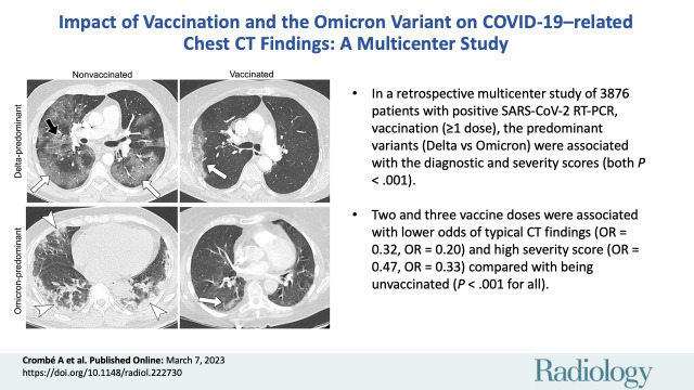
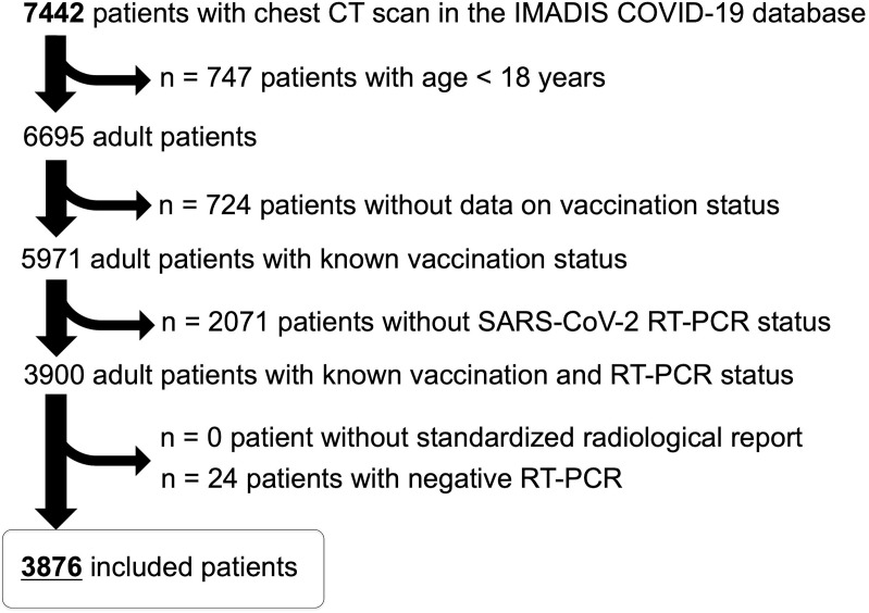
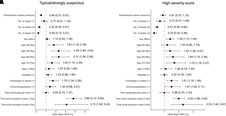
Background The SARS-CoV-2 Omicron variant has a higher infection rate than previous variants but results in less severe disease. However, the effects of Omicron and vaccination on chest CT findings are difficult to evaluate. Purpose To investigate the effect of vaccination status and predominant variant on chest CT findings, diagnostic scores, and severity scores in a multicenter sample of consecutive patients referred to emergency departments for proven COVID-19. Materials and Methods This retrospective multicenter study included adults referred to 93 emergency departments with SARS-CoV-2 infection according to a reverse-transcriptase polymerase chain reaction test and known vaccination status between July 2021 and March 2022. Clinical data and structured chest CT reports, including semiquantitative diagnostic and severity scores following the French Society of Radiology-Thoracic Imaging Society guidelines, were extracted from a teleradiology database. Observations were divided into Delta-predominant, transition, and Omicron-predominant periods. Associations between scores and variant and vaccination status were investigated with χ2 tests and ordinal regressions. Multivariable analyses evaluated the influence of Omicron variant and vaccination status on the diagnostic and severity scores. Results Overall, 3876 patients were included (median age, 68 years [quartile 1 to quartile 3 range, 54-80]; 1695 women). Diagnostic and severity scores were associated with the predominant variant (Delta vs Omicron, χ2 = 112.4 and 33.7, respectively; both P < .001) and vaccination status (χ2 = 243.6 and 210.1; both P < .001) and their interaction (χ2 = 4.3 [P = .04] and 28.7 [P < .001], respectively). In multivariable analyses, Omicron variant was associated with lower odds of typical CT findings than was Delta variant (odds ratio [OR], 0.46; P < .001). Two and three vaccine doses were associated with lower odds of demonstrating typical CT findings (OR, 0.32 and 0.20, respectively; both P < .001) and of having high severity score (OR, 0.47 and 0.33, respectively; both P < .001), compared with unvaccinated patients. Conclusion Both the Omicron variant and vaccination were associated with less typical chest CT manifestations of COVID-19 and lesser extent of disease. © RSNA, 2023 Supplemental material is available for this article. See also the editorial by Yoon and Goo in this issue.
Conflict of interest statement
Figures
![Representative chest CT images of the categories of French Society of
Radiology–Society of Thoracic Imaging scores. (A) Diagnostic scores
are as follows: 1, normal; 2, non–SARS-CoV-2 infection (bacterial
bronchopneumonia [arrowhead]); 3, indeterminate (single ground-glass
opacities [white arrowhead] and contralateral subpleural consolidation
[black arrowhead]); 4, compatible with COVID-19 (bifocal ground-glass
opacities and consolidation [arrowheads]); and 5, typical for COVID-19. (B)
Severity score, depending on the volume of lung parenchyma affected by
COVID-19 (arrowheads indicate subtle focal ground-glass opacities). The
color scheme used for both scores is used in Figures S1–S3. All
images correspond to axial CT pulmonary angiographic images with lung
kernel.](https://cdn.ncbi.nlm.nih.gov/pmc/blobs/836b/10140637/c04a936b83c2/radiol.222730.fig2.jpg)
![Representative examples of patients with different vaccine statuses
during the Delta- and Omicron-predominant periods. (A) Axial CT pulmonary
angiographic scan (with lung kernel settings) in a 65-year-old unvaccinated
male patient during the Delta-predominant period shows CT findings typical
of COVID-19 (ie, bilateral and asymmetric ground-glass opacities [white
arrows] affecting the central and peripheral lung, associated with
reticulations [black arrow] responsible for the crazy-paving pattern). The
severity score assessed based on results of the entire CT pulmonary
angiographic examination was severe extent. (B) Axial CT pulmonary
angiographic scan in a 57-year-old vaccinated male patient (two doses)
during the Delta-predominant period shows chest CT findings compatible with
COVID-19 (ie, single unilateral peripheral ground-glass opacities [arrow])
with a minimal severity score. (C) Axial chest CT scan obtained without
contrast medium in a 69-year-old unvaccinated male patient during the
Omicron-predominant period shows peripheral, bilateral, and asymmetric
consolidations (arrowheads), classified as compatible with COVID-19. The
severity score for the entire chest CT examination was extended. (D) Axial
CT pulmonary angiographic scan in a 70-year-old vaccinated female patient
(three doses) shows a single small peripheral lesion combining ground-glass
opacities and consolidation (arrow) classified as indeterminate, with a
minimal severity score.](https://cdn.ncbi.nlm.nih.gov/pmc/blobs/836b/10140637/0a4d90d119bf/radiol.222730.fig4.jpg)
Comment in
-
Changes in COVID-19 CT Manifestations with Vaccination and the Omicron Variant.Radiology. 2023 May;307(3):e230454. doi: 10.1148/radiol.230454. Epub 2023 Mar 7. Radiology. 2023. PMID: 36880953 Free PMC article. No abstract available.
References
-
- Santé Public France. chiffres clés et évolution de la COVID-19 en France et dans le monde . https://www.santepubliquefrance.fr/dossiers/coronavirus-covid-19/coronav.... Accessed October 22, 2022 .
-
- Simpson S , Kay FU , Abbara S , et al . Radiological Society of North America expert consensus document on reporting chest CT findings related to COVID-19: endorsed by the Society of Thoracic Radiology, the American College of Radiology, and RSNA . Radiol Cardiothorac Imaging 2020. ; 2 ( 2 ): e200152 . - PMC - PubMed
Publication types
MeSH terms
Supplementary concepts
LinkOut - more resources
Full Text Sources
Medical
Miscellaneous
