. 2023 Apr 17;133(8):e166577.
doi: 10.1172/JCI166577.

Callus γδ T cells and microbe-induced intestinal Th17 cells improve fracture healing in mice

Affiliations

Callus γδ T cells and microbe-induced intestinal Th17 cells improve fracture healing in mice

Hamid Y Dar et al. J Clin Invest. .

Abstract

IL-17A (IL-17), a driver of the inflammatory phase of fracture repair, is produced locally by several cell lineages including γδ T cells and Th17 cells. However, the origin of these T cells and their relevance for fracture repair are unknown. Here, we show that fractures rapidly expanded callus γδ T cells, which led to increased gut permeability by promoting systemic inflammation. When the microbiota contained the Th17 cell-inducing taxon segmented filamentous bacteria (SFB), activation of γδ T cells was followed by expansion of intestinal Th17 cells, their migration to the callus, and improved fracture repair. Mechanistically, fractures increased the S1P receptor 1-mediated (S1PR1-mediated) egress of Th17 cells from the intestine and enhanced their homing to the callus through a CCL20-mediated mechanism. Fracture repair was impaired by deletion of γδ T cells, depletion of the microbiome by antibiotics (Abx), blockade of Th17 cell egress from the gut, or Ab neutralization of Th17 cell influx into the callus. These findings demonstrate the relevance of the microbiome and T cell trafficking for fracture repair. Modifications of microbiome composition via Th17 cell-inducing bacteriotherapy and avoidance of broad-spectrum Abx may represent novel therapeutic strategies to optimize fracture healing.

Keywords: Bone Biology; Bone disease; Microbiology; Orthopedics; T cells.


Conflict of interest statement

Conflict of interest: The authors have declared that no conflict of interest exists.

Figures

Figure 1
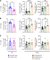
Figure 1. Effects of fractures and Abx-induced microbiota depletion on callus and intestinal inflammatory cytokine transcripts and on the relative number of callus and intestinal γδ T cells and Th17 cells.
(A) Effects of fractures on the levels of Il17a, Tnf, Il1b, and Il6 transcripts in the callus of SFB+ and SFB JAX mice. (B) Effects of fractures on the levels of Il17a, Tnf, Il1b, and Il6 transcripts in the SI of SFB+ and SFB JAX mice. (C) Effects of fractures on the relative frequency of γδ T cells (CD3ε+CD45+TCRγδ+) and Th17 cells (TCRβ+CD45+CD4+IL-17A+) in the callus and PPs of SFB+ and SFB JAX mice. Femoral fractures were induced in 12-week-old female SFB+ JAX and SFB JAX mice. Mice were treated or not with broad-spectrum Abx starting 1 week before fracture surgery. PP and callus cells were recovered daily for 3 days after fracture surgery and analyzed by flow cytometry. Time 0 indicates intact bone. n = 5 mice/group. Data are expressed as the mean ± SEM. All data were normally distributed according to the Shapiro-Wilk normality test and analyzed by 2-way ANOVA with post hoc Bonferroni correction for multiple comparisons. *P < 0.05, **P < 0.01, ***P < 0.001, and ****P < 0.0001 compared with the indicated group. Nonsignificant comparisons are not shown.
Figure 2
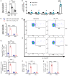
Figure 2. Effects of fractures and Abx-induced microbiota depletion on callus and intestinal γδ T cells and Th17 cells, and gut permeability in WT and γδ T cell–/– mice.
Femoral fractures were induced in 12-week-old female SFB+ JAX mice, SFB JAX mice, and SFB+ Tcrd–/– mice, a strain lacking γδ T cells. SFB+ JAX mice and SFB JAX mice were treated or not with broad-spectrum Abx starting 1 week before fracture surgery. Frequencies of (A) callus γδ T cells (CD3ε+CD45+TCRγδ+ cells), (B) PP γδ T cells, (C) callus Th17 cells (TCRβ+CD45+CD4+IL-17A+ cells), and (D) PP Th17 cells. (E) Callus and PP Th17 cells in Tcrd–/– mice. (F) Gut permeability in SFB+ WT mice and SFB+ Tcrd–/– mice. Gut permeability was assessed by serum LPS levels and FITC-dextran absorption. n = 5 mice/group. Data are expressed as the mean ± SEM. All data were normally distributed according to the Shapiro-Wilk normality test and analyzed by 2-way ANOVA and with post hoc Bonferroni correction for multiple comparisons. **P < 0.01, ***P < 0.001, and ****P < 0.0001 compared with the indicated group (AE) or compared with intact bone (F). Nonsignificant comparisons are not shown.
Figure 3
Figure 3. Fractures increase homing to the callus of intestinal αβ T cells and Th17 cells but not γδ T cells.
Femoral fractures were induced in 12-week-old male SFB+ Kaede mice. After 2 days, 4 PPs were surgically exposed and illuminated with a near-UV light for 2 minutes. After 1 day, mice were sacrificed, and the frequency of PPs and callus red-fluorescing αβ T cells, Th17 cells, and γδ T cells was determined by flow cytometry. PP red-fluorescing T cells were counted in PPs from mice with fractures and PPs from uninjured control mice. Callus red-fluorescing T cells were counted in the callus tissue of fractured femurs, BM from the contralateral uninjured femur, and BM from uninjured mice. (A) Relative frequency of PP αβ T cells and relative and absolute frequency of callus αβ T cells. (B) Relative frequency of PP Th17 cells and relative and absolute frequency of callus Th17 cells. (C) Relative frequency of PP γδ T cells and relative and absolute frequency of callus γδ T cells. n = 6 mice/group. Data are expressed as the mean ± SEM. All data were normally distributed according to the Shapiro-Wilk normality test and analyzed by 2-way ANOVA with post hoc Bonferroni correction for multiple comparisons (callus panels), or by unpaired, 2-tailed t test (PP panels). *P < 0.05, ***P < 0.001, and ****P < 0.0001 compared with the indicated group. Nonsignificant comparisons are not shown.
Figure 4
Figure 4. Fractures increase the tropism of Th17 cells to the callus via a TNF-dependent mechanism.
(A) Callus Ccl20 transcript levels at days 3 and 7 PF in SFB+ TAC mice. (B) Ccl20 transcript levels in purified callus cells at day 3 PF. (C) Callus Ccl20 transcript levels in SFB+ WT and Tnf–/– mice at day 3 PF. (D and E) Relative and absolute frequencies of EGFP+ Th17 cells in the callus of SFB+ WT and Tnf–/– mice subjected to fracture 2 days before adoptive transfer of IL-17A-EGFP+ cells. (F) Relative and absolute frequencies of callus Th17 cells in SFB+ WT mice and Tnf–/– mice. (G) Relative frequency of callus Vβ14+ Th17 cells in SFB+ WT and Tnf–/– mice. n = 5–6 mice/group. Data are expressed as the mean ± SEM. All data were normally distributed according to the Shapiro-Wilk normality test and analyzed by 2-way ANOVA with post hoc Bonferroni correction for multiple comparisons. **P < 0.01, ***P < 0.001, and ****P < 0.0001 compared with the indicated group. #P < 0.001 compared with day 3 PF SCs. Nonsignificant comparisons are not shown.
Figure 5
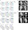
Figure 5. γδ T cells and Th17 cells accelerate fracture healing.
Femoral fractures were induced in 12-week-old female SFB+ and SFB JAX mice, and SFB+ Tcrd–/– mice, a strain lacking γδ T cells. (A) Callus μCT measurements at days 14 and 21 PF in WT and Tcrd–/– mice. (B) Representative images of 3D μCT reconstructions of fracture callus from WT and Tcrd–/– mice. (C) Callus μCT measurements at days 14 and 21 in SFB+ and SFB JAX mice. (D) Representative images of 3D μCT reconstructions of fracture callus from SFB+ and SFB JAX mice at days 14 and 21. (E) Torsion stiffness, yield torque, and ultimate torque at day 35 PF were determined by static torsion-to-failure testing of excised femora. Data are expressed as the mean ± SEM. All data were normally distributed according to the Shapiro-Wilk normality test and analyzed by unpaired t test. *P < 0.05, **P < 0.01, and ***P < 0.001 compared with the indicated group. Nonsignificant comparisons are not shown.
Figure 6
Figure 6. Blockade of Th17 cell egress from the intestine prevents the fracture-induced increase in peripheral blood and callus Th17 cells and impairs fracture healing.
(A) Relative frequency of PP, peripheral blood (PB), and callus Th17 cells at days 3 and 7 PF in mice treated with the S1PR1 blocker FTY720. (B) Effects of fractures on the transcript levels of Il17a in the SI and callus in mice treated with FTY720. (C and D) Callus μCT measurements at day 14 PF. Mice were treated with FTY720 for 2 weeks starting 1 week before fracture surgery. n = 5–6 mice/group. Data are expressed as the mean ± SEM. All data were normally distributed according to the Shapiro-Wilk normality test and analyzed by 2-way ANOVA with post hoc Bonferroni correction for multiple comparisons, or by unpaired, 2-tailed t tests. *P < 0.05, ***P < 0.001, and ****P < 0.0001, compared with the indicated group. Nonsignificant comparisons are not shown.
Figure 7
Figure 7. Blockade of Th17 cell influx into callus by treatment with anti CCL20 Ab prevents the fracture-induced increase in peripheral blood and callus Th17 cells and impairs fracture healing.
(A) Relative frequency of PP, peripheral blood, and callus Th17 cells at days 3 and 7 PF in mice treated with anti-CCL20 Ab. (B) Effect of fractures on transcript levels of Il17a in the SI and callus in mice treated with anti-CCL20 Ab. (C and D) Callus μCT measurements at day 14 PF. Mice were treated with anti-CCL20 Ab or irrelevant (Irr.) Ab 1 day before surgery and every other day for 7 days. n = 5–6 mice/group. Data are expressed as the mean ± SEM. All data were normally distributed according to the Shapiro-Wilk normality test and analyzed by 2-way ANOVA with post hoc Bonferroni correction for multiple comparisons. *P < 0.05, ***P < 0.001, and ****P < 0.0001, compared with the indicated group. Nonsignificant comparisons are not shown.

Comment in

References

    1. Baht GS, et al. The role of the immune cells in fracture healing. Curr Osteoporos Rep. 2018;16(2):138–145. doi: 10.1007/s11914-018-0423-2. - DOI - PMC - PubMed
    1. Einhorn TA, Gerstenfeld LC. Fracture healing: mechanisms and interventions. Nat Rev Rheumatol. 2015;11(1):45–54. doi: 10.1038/nrrheum.2014.164. - DOI - PMC - PubMed
    1. Marsell R, Einhorn TA. The biology of fracture healing. Injury. 2011;42(6):551–555. doi: 10.1016/j.injury.2011.03.031. - DOI - PMC - PubMed
    1. Colnot C, et al. Analyzing the cellular contribution of bone marrow to fracture healing using bone marrow transplantation in mice. Biochem Biophys Res Commun. 2006;350(3):557–561. doi: 10.1016/j.bbrc.2006.09.079. - DOI - PubMed
    1. Nam D, et al. T-lymphocytes enable osteoblast maturation via IL-17F during the early phase of fracture repair. PLoS One. 2012;7(6):e40044. doi: 10.1371/journal.pone.0040044. - DOI - PMC - PubMed

Publication types

Substances