. 2023 Jun;531(8):838-852.
doi: 10.1002/cne.25467. Epub 2023 Mar 7.

Early development of respiratory motor circuits in larval zebrafish (Danio rerio)

Affiliations

Early development of respiratory motor circuits in larval zebrafish (Danio rerio)

Kimberly L McArthur et al. J Comp Neurol. 2023 Jun.

Abstract

Rhythm-generating circuits in the vertebrate hindbrain form synaptic connections with cranial and spinal motor neurons, to generate coordinated, patterned respiratory behaviors. Zebrafish provide a uniquely tractable model system to investigate the earliest stages in respiratory motor circuit development in vivo. In larval zebrafish, respiratory behaviors are carried out by muscles innervated by cranial motor neurons-including the facial branchiomotor neurons (FBMNs), which innervate muscles that move the jaw, buccal cavity, and operculum. However, it is unclear when FBMNs first receive functional synaptic input from respiratory pattern-generating neurons, and how the functional output of the respiratory motor circuit changes across larval development. In the current study, we used behavior and calcium imaging to determine how early FBMNs receive functional synaptic inputs from respiratory pattern-generating networks in larval zebrafish. Zebrafish exhibited patterned operculum movements by 3 days postfertilization (dpf), though this behavior became more consistent at 4 and 5 dpf. Also by 3dpf, FBMNs fell into two distinct categories ("rhythmic" and "nonrhythmic"), based on patterns of neural activity. These two neuron categories were arranged differently along the dorsoventral axis, demonstrating that FBMNs have already established dorsoventral topography by 3 dpf. Finally, operculum movements were coordinated with pectoral fin movements at 3 dpf, indicating that the operculum behavioral pattern was driven by synaptic input. Taken together, this evidence suggests that FBMNs begin to receive initial synaptic input from a functional respiratory central pattern generator at or prior to 3 dpf. Future studies will use this model to study mechanisms of normal and abnormal respiratory circuit development.

Keywords: facial branchiomotor neurons; motor circuits; respiratory behavior; respiratory circuits; respiratory development; zebrafish.


Conflict of interest statement

CONFLICT OF INTEREST DISCLOSURE

The authors declare no conflicts of interest.

Figures

Figure 1.
Figure 1.. High-speed behavioral imaging demonstrates that larval zebrafish can produce bouts of patterned operculum movements at 3dpf.
(a) At 3dpf, some fish (n = 2 of 13) exhibited irregular operculum movements of varying amplitude and frequency, without a clear pattern. (b) Most fish at 3dpf (n = 11 of 13) exhibited bouts of rhythmic operculum movement, followed by a large burst of operculum movement associated with attempted whole-body movement (indicated by arrows), followed by a pause before rhythmic movement resumed. In both (a) and (b), a microscopic image of the operculum is shown in the upper left, and a representative trace of dorsoventral operculum position over time is shown in the upper right. The location of the landmark used to track movements in this subject is indicated by an asterisk. A histogram (bottom) shows the distribution of inter-movement intervals for this subject across trials (excluding intervals during and immediately following attempted whole-body movements). The histogram has been divided into smaller (<5s, left) and larger (>5s, right) intervals.
Figure 2.
Figure 2.. Operculum behavior becomes more consistently patterned at 4dpf and 5dpf.
At both (a) 4dpf (n = 15) and (b) 5dpf (n = 15), all subjects produced bouts of rhythmic operculum movement, followed by a large burst of operculum movement associated with attempted whole-body movement (indicated by arrows), followed by a pause before rhy-thmic movement resumed. Note that rhythmic movements could occur as couplets, as shown for the example in (b). In both (a) and (b), a microscopic image of the operculum is shown in the upper left, and a representative trace of dorsoventral operculum position over time is shown in the upper right. The location of the landmark used to track movements in this subject is indicated by an asterisk. A histogram (bottom) shows the distribution of inter-movement intervals for this subject across trials (excluding intervals during and immediately following attempted whole-body movements). The histogram has been divided into smaller (<5s, left) and larger (>5s, right) intervals.
Figure 3.
Figure 3.. Bouts of patterned operculum movements at 3dpf have similar temporal properties overall to those observed at 4dpf and 5dpf.
Histograms show the distribution of operculum inter-movement intervals across subjects at (a) 3dpf (n = 13; 4842 total intervals), (b) 4dpf (n = 15; 13657 total intervals), and (c) 5dpf (n = 15; 11637 total intervals). The number of total intervals falling within a particular range (in seconds) is shown as a percentage of the total number of intervals in that age group. Intervals during and immediately following attempted whole-body movements were excluded from this analysis, to focus on the rhythmic component of the behavior. Fish produced short-interval operculum movements (<1s) at all three ages, though fish exhibited many more long (>5s) intervals at 3dpf than at 4 or 5dpf.
Figure 4.
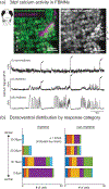
Figure 4.. Calcium imaging demonstrates that FBMNs exhibit both rhythmic and non-rhythmic activity at 3dpf, and reveals dorsoventral topography by activity pattern.
(a) At 3dpf, some FBMNs (i) exhibit only large, infrequent (“non-rhythmic”) calcium transients associated with attempted whole-body movements (indicated by arrows), while other FBMNs (ii and iii) exhibit bouts of additional burst activity (“rhythmic”). Sampling interval = 295ms. (b) At 3dpf, rhythmic FBMNs are located almost exclusively in the ventral half of the facial motor nucleus (0–19μm from the ventral-most FBMN, for each fish), while non-rhythmic FBMNs are found throughout its dorsoventral extent. Response category (rhythmic vs. non-rhythmic) is a significant predictor of dorsoventral position (n=6 fish; mixed effects linear model: p = 0.0035). Bars of the same color indicate FBMNs imaged in the same fish. Dorsoventral position is measured with respect to the approximate plane of the bilateral facial motor nerve within the hindbrain.
Figure 5.
Figure 5.. Operculum movements are coordinated with pectoral fin movements at both 3dpf and 5dpf.
(a) At 3dpf, all subjects (n = 9) produced bouts of high-frequency (10–35Hz) pectoral fin movements, typically accompanied by one or more operculum movements. (b) At 5dpf, all subjects (n = 8) produced shorter bouts of high-frequency (10–50Hz) pectoral fin movements, almost always accompanied by one or more operculum movements. Operculum movements rarely occurred in the absence of pectoral fin movement, at either age. In both (a) and (b), a lateral view microscopic image of the subject is shown in the upper left, accompanied by a line drawing that highlights key anatomical features used to track fin and operculum movements. The plots underneath the images show the peak displacement times for the operculum and pectoral fin during a 30s period, represented as vertical lines.
Figure 6.
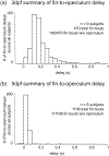
Figure 6.. Operculum-fin coordination becomes tighter and less variable between 3dpf and 5dpf.
Histograms show the distribution of delays between pectoral fin and operculum movements at (a) 3dpf (n = 9; 325 fin-operculum delays shown, from 470 total fin bouts – 145/470 fin bouts occurred without operculum movement) and (b) 5dpf (n = 8; 1107 total fin-operculum delays shown, from 1108 total fin bouts – 1/1108 fin bouts occurred without operculum movement). Fin-to-operculum delay was calculated as the difference in the peak displacement time of the first pectoral fin and operculum movements, within a single pectoral fin bout. A positive value indicates that the fin reached peak displacement before the operculum, and a negative value indicates that the operculum reached peak displacement before the fin. Note that most delay values are positive, indicating that the fin typically leads the operculum. This delay was longer overall at 3dpf (median delay = 0.17s) than 5dpf (median delay = 0.03s), confirmed by statistical comparison (mixed effects linear model: p < 0.0001).

References

    1. Asante E, Hummel D, Gurung S, Kassim YM, Al-Shakarji N, Palaniappan K, Sittaramane V, Chandrasekhar A. 2021. Defective neuronal positioning correlates with aberrant motor circuit function in zebrafish. Front Neural Circuits 15:690475. DOI: 10.3389/fncir.2021.690475. - DOI - PMC - PubMed
    1. Bass AH, Baker R. 1997. Phenotypic specification of hindbrain rhombomeres and the origins of rhythmic circuits in vertebrates. Brain Behav Evol 50Suppl 1):3–16. DOI: 10.1159/000113351 - DOI - PMC - PubMed
    1. Bongianni F, Mutolo D, Cinelli E, Pantaleo T. 2014. Neural mechanisms underlying respiratory rhythm generation in the lamprey. Respir Physiol Neurobiol 224:17–26. DOI: 10.1016/j.resp.2014.09.003 - DOI - PubMed
    1. Camp AL, Brainerd EL. 2015. Reevaluating musculoskeletal linkages in suction-feeding fishes with X-ray reconstruction of moving morphology (XROMM). Integ Comp Biol 55(1):36–47. DOI: 10.1093/icb/icv034 - DOI - PubMed
    1. Chandrasekhar A, Moens CB, Warren JT Jr, Kimmel CB, Kuwada JY. 1997. Development of branchiomotor neurons in zebrafish. Development 124(13):2633–44. DOI: 10.1242/dev.124.13.2633 - DOI - PubMed

Publication types

Substances