Review
. 2023 Mar 8;14(3):186.
doi: 10.1038/s41419-023-05708-0.

Iron metabolism and ferroptosis in type 2 diabetes mellitus and complications: mechanisms and therapeutic opportunities

Affiliations
Review

Iron metabolism and ferroptosis in type 2 diabetes mellitus and complications: mechanisms and therapeutic opportunities

Runyu Miao et al. Cell Death Dis. .

Abstract

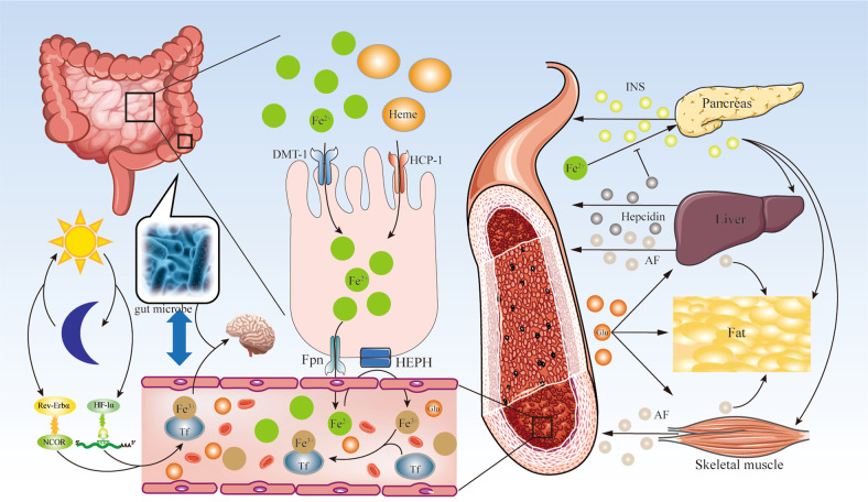
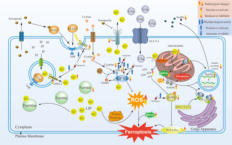
The maintenance of iron homeostasis is essential for proper endocrine function. A growing body of evidence suggests that iron imbalance is a key factor in the development of several endocrine diseases. Nowadays, ferroptosis, an iron-dependent form of regulated cell death, has become increasingly recognized as an important process to mediate the pathogenesis and progression of type 2 diabetes mellitus (T2DM). It has been shown that ferroptosis in pancreas β cells leads to decreased insulin secretion; and ferroptosis in the liver, fat, and muscle induces insulin resistance. Understanding the mechanisms concerning the regulation of iron metabolism and ferroptosis in T2DM may lead to improved disease management. In this review, we summarized the connection between the metabolic pathways and molecular mechanisms of iron metabolism and ferroptosis in T2DM. Additionally, we discuss the potential targets and pathways concerning ferroptosis in treating T2DM and analysis the current limitations and future directions concerning these novel T2DM treatment targets.


Conflict of interest statement

The authors declare no competing interests.

Figures

Fig. 1
Fig. 1. Flow chart of the study.
Iron metabolism. Iron is absorbed by intestinal cells in the form of free divalent iron (Fe2+) or heme iron (Heme) in the intestine. Fe2+ is absorbed by intestinal cells through divergent metal transporter 1 (DMT-1). Heme iron is transported to intestinal cells through heme carrier protein 1 (HCP-1). Fe2+ is released into the blood capillary through ferroportin (Fpn), oxidized into free ferric iron (Fe3+) by hephaestin (HEPN), combined with transferrin (Tf) in the circulation, and transported to organs and tissues. Fe3+ enters the pancreas, liver, fat, and skeletal muscle to regulate blood glucose (Glu) homeostasis. Pancreas β Cells release insulin (INS) in response to the stimulation of Glu. INS affects the liver, fat, and skeletal muscles. The liver and skeletal muscles release adipose factor (AF), which regulates adipose tissue metabolism. On the other hand, iron metabolism also influences the composition of the gut microbiota, which may affect cognitive through the gut-brain axis. Iron affects circadian glucose metabolism via the regulation of the interaction of nuclear receptor subfamily 1 group d member 1 (Rev-Erbα) with its co-suppressor, nuclear receptor corepressor 1 (NCOR). iron also participates in the regulation of β-cell function mediated by HIF-1α in circadian rhythms.
Fig. 2
Fig. 2. Iron metabolism in pancreatic β cells.
Iron metabolism in regulating insulin secretion in pancreatic β cells. Fe3+ combined with transferrin (Tf) into Tf-Fe2+ in the circulation. Tf-Fe2+ binds to the transferrin receptor (TrfR) on the cell surface, and the receptor complex is endocytosed with the divergent metal transporter 1 (DMT-1). Inside the endosome, Fe3+ is reduced to Fe2+ and released into the labile iron pool (LIP). Ferritin combines with Fe2+ in LIP to regulate the concentration of Fe2+ in cells. In addition, Fe2+ is discharged from cells through ferroportin (Fpn). The pancreas β cells, and hepatocyte can release hepcidin, which can induce Tf internalized and inhibit the activity of Fpn. Glucose (Glu) enters the pancreas β cells via glucose transporter 2 (GLUT-2) and performs glycolysis before entering the mitochondria, which leads to increased ATP production. Fe2+ promotes the production of reactive oxygen species (ROS) through the Fenton reaction. Iron exchange in the mitochondria is mediated by DMT-1 and classical mitoferrins (Mfrn) 1 and 2, which can be incorporated into the electron transport chain and produce more ATP under the stimulation of glucose. Fe2+ participates in Fe-S cluster biosynthesis in mitochondria. The Fe-S cluster promotes Cdkal1 catalytic metabolism of t6A37 in tRNALysUUU to ms2t6A37 and enables the normal processing of proinsulin into insulin.
Fig. 3
Fig. 3. Pharmacological target for treating ferroptosis in pancreatic β cells.
Ferroptosis in pancreatic β cells and pharmacological target mechanisms of different drugs. Pancreatic β cells express low levels of antioxidant enzymes, such as superoxide dismutase (SOD), glutathione (GSH) peroxidase, and glutathione peroxidase 4 (Gpx4). The Fe2+ in the labile iron pool (LIP) promotes reactive oxygen species synthesis through the Fenton reaction, leading to the accumulation of reactive oxygen species (ROS). External factors will cause mitochondria damage and produce an excess of mitochondrial ROS (MtROS). ROS and MtROS lead to ROS-dependent autophagy and ferroptosis, and cause intracellular iron increased. Iron in mitochondria accumulation will cause the lack of Fe-S clusters, which could lead to ROS increase in the mitochondria. The lack of Fe-S cluster and the increase of ROS will reduce the synthesis and secretion of insulin. Metformin, Quercetin, Melatonin, and Vitamin D effect on different targets to reduce the possibility of ferroptosis in pancreatic β cells.

References

    1. Forbes A. Iron and parenteral nutrition. Gastroenterology. 2009;137(5 Suppl):S47–S54. doi: 10.1053/j.gastro.2009.08.013. - DOI - PubMed
    1. Prá D, Franke SI, Henriques JA, Fenech M. Iron and genome stability: an update. Mutat Res. 2012;733:92–9. doi: 10.1016/j.mrfmmm.2012.02.001. - DOI - PubMed
    1. Hentze MW, Muckenthaler MU, Galy B, Camaschella C. Two to tango: regulation of mammalian iron metabolism. Cell. 2010;142:24–38. doi: 10.1016/j.cell.2010.06.028. - DOI - PubMed
    1. Soe-Lin S, Apte SS, Andriopoulos B, Jr, Andrews MC, Schranzhofer M, Kahawita T, et al. Nramp1 promotes efficient macrophage recycling of iron following erythrophagocytosis in vivo. Proc Natl Acad Sci USA. 2009;106:5960–5. doi: 10.1073/pnas.0900808106. - DOI - PMC - PubMed
    1. Jordan JB, Poppe L, Haniu M, Arvedson T, Syed R, Li V, et al. Hepcidin revisited, disulfide connectivity, dynamics, and structure. J Biol Chem. 2009;284:24155–67. doi: 10.1074/jbc.M109.017764. - DOI - PMC - PubMed

Publication types