. 2023 Apr 11;147(15):1162-1179.
doi: 10.1161/CIRCULATIONAHA.122.063073. Epub 2023 Mar 8.

Nitrative Modification of Caveolin-3: A Novel Mechanism of Cardiac Insulin Resistance and a Potential Therapeutic Target Against Ischemic Heart Failure in Prediabetic Animals

Affiliations

Nitrative Modification of Caveolin-3: A Novel Mechanism of Cardiac Insulin Resistance and a Potential Therapeutic Target Against Ischemic Heart Failure in Prediabetic Animals

Zhijun Meng et al. Circulation. .

Erratum in

Abstract

Background: Myocardial insulin resistance is a hallmark of diabetic cardiac injury. However, the underlying molecular mechanisms remain unclear. Recent studies demonstrate that the diabetic heart is resistant to other cardioprotective interventions, including adiponectin and preconditioning. The "universal" resistance to multiple therapeutic interventions suggests impairment of the requisite molecule(s) involved in broad prosurvival signaling cascades. Cav (Caveolin) is a scaffolding protein coordinating transmembrane signaling transduction. However, the role of Cav3 in diabetic impairment of cardiac protective signaling and diabetic ischemic heart failure is unknown.

Methods: Wild-type and gene-manipulated mice were fed a normal diet or high-fat diet for 2 to 12 weeks and subjected to myocardial ischemia and reperfusion. Insulin cardioprotection was determined.

Results: Compared with the normal diet group, the cardioprotective effect of insulin was significantly blunted as early as 4 weeks of high-fat diet feeding (prediabetes), a time point where expression levels of insulin-signaling molecules remained unchanged. However, Cav3/insulin receptor-β complex formation was significantly reduced. Among multiple posttranslational modifications altering protein/protein interaction, Cav3 (not insulin receptor-β) tyrosine nitration is prominent in the prediabetic heart. Treatment of cardiomyocytes with 5-amino-3-(4-morpholinyl)-1,2,3-oxadiazolium chloride reduced the signalsome complex and blocked insulin transmembrane signaling. Mass spectrometry identified Tyr73 as the Cav3 nitration site. Phenylalanine substitution of Tyr73 (Cav3Y73F) abolished 5-amino-3-(4-morpholinyl)-1,2,3-oxadiazolium chloride-induced Cav3 nitration, restored Cav3/insulin receptor-β complex, and rescued insulin transmembrane signaling. It is most important that adeno-associated virus 9-mediated cardiomyocyte-specific Cav3Y73F reexpression blocked high-fat diet-induced Cav3 nitration, preserved Cav3 signalsome integrity, restored transmembrane signaling, and rescued insulin-protective action against ischemic heart failure. Last, diabetic nitrative modification of Cav3 at Tyr73 also reduced Cav3/AdipoR1 complex formation and blocked adiponectin cardioprotective signaling.

Conclusions: Nitration of Cav3 at Tyr73 and resultant signal complex dissociation results in cardiac insulin/adiponectin resistance in the prediabetic heart, contributing to ischemic heart failure progression. Early interventions preserving Cav3-centered signalsome integrity is an effective novel strategy against diabetic exacerbation of ischemic heart failure.

Keywords: caveolin 3; diabetes mellitus; protein processing, posttranslational; reperfusion injury.


Conflict of interest statement

Disclosures

The authors declare no competing interests, financial or otherwise.

Figures
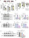
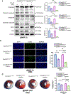
Figure 1.
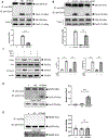
Figure 1.. Impairment of cardioprotective response and transmembrane signaling occurred long before HFD-induced type 2 diabetes.
A. Infarct-sparing effect of insulin was detected by Evans blue-TTC staining in ND and HFD (4- and 12-weeks) mice. B. Global longitudinal strain (GLS) determined by echocardiography in ND and HFD (4- and 12-weeks) mice. n = 18–20/group. *p < 0.05, **p < 0.01 vs. vehicle, respectively. C. Transmembrane signaling was determined by Akt phosphorylation and Glut4 membrane translocation 30 min after insulin treatment in adult cardiomyocytes after HFD 4 weeks. n = 5–6/group, *p < 0.05, **p < 0.01 vs. vehicle, respectively. D. IRβ and IRS-1 expression in ND and HFD (4- and 12-weeks) heart. n = 6/group, **p < 0.01 vs. ND group. E. Cav3 expression in ND and HFD (2–12 weeks) heart. n = 5–6/group, **p < 0.01 vs. ND group. ND: normal diet; HFD: high-fat diet; MI/R: myocardial ischemia/reperfusion; IRβ: insulin receptor β; IRS-1: Insulin receptor substrate-1.
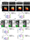
Figure 2.
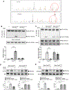
Figure 2.. Cav3-centered signalsomes were dissociated in the prediabetic heart, a pathologic effect phenocopied by SIN1.
A. Detection of Cav3/IRβ complex interaction in ND or HFD (4 weeks) heart samples. n = 6/group, **p < 0.01 vs. ND. B. Detection of Cav3/IRβ association in SIN1-treated adult cardiomyocytes in the presence and absence of SOD. n = 6/group, **p < 0.01 vs. SIN1 group. C. Insulin-induced Akt phosphorylation and Glut4 membrane translocation in adult cardiomyocytes were blocked by SIN1, an effect rescued by SOD. n = 6/group, **p < 0.01 vs. vehicle, ##p < 0.01 vs. SIN1 without SOD. D/E. Cav3, not IRβ, was significantly nitrated 4 weeks post-HFD. The level of nitrated Cav3/total Cav3 was about 38.77±7.9%. n = 6/group. **p < 0.01 vs. ND. SIN1: 5-amino-3-(4-morpholinyl)-1,2,3-oxadiazolium chloride; SOD: Superoxide Dismutase.
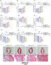
Figure 3.
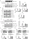
Figure 3.. Identifying Cav3Y73 as the responsible site for nitrative dissociation of Cav3 signalsomes.
A. MS analysis identified Tyr73 nitration in prediabetic but not in normal control heart. B. SIN1-induced Cav3 nitration was detected in Cav3KO neonatal mouse cardiomyocytes re-expressing WT Cav3 (Adv-Cav3WT) but not in Cav3KO neonatal mouse cardiomyocytes re-expressing Tyr73 mutated Cav3 (Adv-Cav3Y73F). n = 6/group. **p < 0.01 vs. Adv-Cav3WT. C. SIN1 caused Cav3/IRβ complex dissociation in Adv-Cav3WT neonatal mouse cardiomyocytes, an effect blocked in Adv-Cav3Y73F neonatal mouse cardiomyocytes. n = 6/group, **p < 0.01 vs. Adv-Cav3WT. D/E. 48 hours HG/HL culture significantly attenuated insulin transmembrane signaling in Adv-Cav3WT neonatal mouse cardiomyocytes (attenuated Akt phosphorylation and reduced Glut4 membrane translocation). Re-expressing Cav3Y73F in Cav3KO neonatal mouse cardiomyocytes rescued insulin-induced Akt phosphorylation (D) and Glut4 membrane translocation (E) after HG/HL treatment. n = 6/group. **p < 0.01 vs. Adv-Cav3WT treated with vehicle. ##P<0.01 vs. Adv-Cav3Y73F treated with vehicle.
Figure 4.
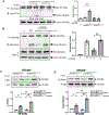
Figure 4.. Tyr73 nitration dissociates Cav3/AdipoR1 and impairs APN transmembrane signaling. A/B.
Detection of Cav3/AdipoR1 association in SIN1-treated adult cardiomyocytes in the presence and absence of SOD. n = 6/group, **p < 0.01 vs. SIN1 group. B. APN-induced AMPK, Akt and ERK1/2 phosphorylation in adult cardiomyocytes were blocked by SIN1, an effect rescued by SOD. n = 6/group, **p < 0.01 vs. vehicle, ##p < 0.01 vs. SIN1 without SOD. C. SIN1 caused Cav3/AdipoR1 complex dissociation in Adv-Cav3WT neonatal mouse cardiomyocytes, an effect blocked in Adv-Cav3Y73F neonatal mouse cardiomyocytes. n = 6/group, **p < 0.01 vs. Adv-Cav3WT. D/E. 48 hours HG/HL culture significantly attenuated APN transmembrane signaling in Adv-Cav3WT neonatal mouse cardiomyocytes (attenuated AMPK and Akt phosphorylation). Re-expressing Cav3Y73F in Cav3KO neonatal mouse cardiomyocytes rescued APN-induced AMPK (D) and Akt phosphorylation (E) after HG/HL treatment. n = 6/group. **p < 0.01 vs. Adv-Cav3WT treated with vehicle. ##P<0.01 vs. Adv-Cav3Y73F treated with vehicle.
Figure 5.
Figure 5.. Re-expressing Cav3Y73F in Cav3KO mice (Cav3KOCav3Y73F) blocked HFD-induced Cav3 nitration, preserved Cav3 signalsome integrity, and rescued transmembrane signaling.
A. HFD causes significant Cav3 nitration in cardiac tissue from Cav3KO mice re-expressing Cav3WT (Cav3KOCav3WT), a pathologic effect blocked in Cav3KOCav3Y73F heart. The level of nitrated Cav3/total Cav3 was about 29.9±4.2% n = 6/group. **p < 0.01 vs. Cav3KOCav3WT. B. HFD dissociated Cav3/IRβ complex in cardiac tissue from Cav3KOCav3WT mice, a pathologic effect blocked in Cav3KOCav3Y73F heart. n = 6/group. **p < 0.01 vs. Cav3KOCav3WT. C/D. Insulin transmembrane signaling (Akt phosphorylation and Glut4 membrane translocation) was significantly improved in cardiomyocytes isolated from Cav3KOCav3Y73F heart subjected to HFD. n = 6/group, *p < 0.05 vs. Cav3KOCav3WT treated with vehicle; **p < 0.01 vs. Cav3KOCav3Y73F treated with vehicle; ##P<0.01 vs. Cav3KOCav3WT treated with insulin.
Figure 6.
Figure 6.. Anti-apoptotic and infarct sparing effect of insulin were enhanced in Cav3KOCav3Y73F mice. A/B.
Prediabetic mice (4-week HFD) were subjected to acute MI/R (60-minute MI followed by 3 hours reperfusion). Mice were treated with vehicle or insulin 10 minutes before reperfusion. Apoptosis was determined by caspase 3 activation, caspase 9 activation, Bax/Bcl2 ratio and TUNEL staining. n = 6–10/group. *p < 0.05 vs. Cav3KOCav3WT treated with vehicle; **p < 0.01 vs. Cav3KOCav3Y73F treated with vehicle; #P<0.05 & ##P<0.01 vs. Cav3KOCav3WT treated with insulin. C. Pre-diabetic mice were subjected to 60-minute MI and treated with vehicle or insulin 10 minutes before reperfusion. Infarct size was determined 24 hours after reperfusion by Evans blue/TTC double stain. n = 8–10/group. *p < 0.05 vs. Cav3KOCav3WT treated with vehicle; **p < 0.01 vs. Cav3Cav3Y73F treated with vehicle; #P<0.05 vs. Cav3KOCav3WT treated with insulin.
Figure 7.
Figure 7.. The acute cardioprotective effect of insulin was improved in Cav3KOCav3Y73F mice.
Prediabetic mice were subjected to 60-minute MI and treated with vehicle or insulin 10 minutes before reperfusion. Cardiac function was determined 24 hours after reperfusion by echocardiography. A. Typical images of three-dimensional regional wall velocity diagrams of radial strain showing contraction (orange/positive values) of 3 consecutive cardiac cycles. Vector diagrams showing the direction and magnitude of endocardial contraction at systole. B. Compared to Cav3KOCav3WT mice, insulin administration had a greater improvement effect upon LVEF and FS in Cav3KOCav3Y73F mice. n = 18–20 mice/group. *p < 0.05 vs. Cav3KOCav3WT treated with vehicle; **p < 0.01 vs. Cav3Cav3Y73F treated with vehicle; ##P<0.01 vs. Cav3KOCav3WT treated with insulin. C. Representative images of speckle-tracking analysis in the long-axis B-mode and radial segmental synchronicity of the LV. D. Global strain and strain rate measured in the radial and longitudinal axes across the LV endocardium. Compared to Cav3KOCav3WT mice, insulin administration had a greater improvement effect on the global radial strain, radial strain rate, global longitudinal strain, and longitudinal strain rate in Cav3KOCav3Y73F mice. n = 18–20 mice/group. *p < 0.05 vs. Cav3KOCav3WT treated with vehicle; **p < 0.01 vs. Cav3Cav3Y73F treated with vehicle; #P<0.05 vs. Cav3KOCav3WT treated with insulin.
Figure 8.
Figure 8.. Chronic cardioprotective and anti-remodeling effects of insulin were preserved in Cav3KOCav3Y73F mice.
Prediabetic mice were subjected to 60-minute MI and treated with vehicle or insulin (bolus administration 10 minutes before reperfusion followed by continuous administration for 4 weeks via an osmatic pump). A-F. Insulin administration failed to improve cardiac function 4 weeks after reperfusion in Cav3KOCav3WT mice fed with HFD. In contrast, insulin significantly improved cardiac function in HFD Cav3KOCav3Y73F mice. n = 18–20/group. *p < 0.05 or **p < 0.01 vs. Cav3KOCav3WT treated with vehicle. #p < 0.05 or ##p < 0.01 vs. Cav3KOCav3Y73F treated with vehicle. G/H. Insulin treatment significantly reduced ratios of heart weight/body weight and lung weight/body weight in Cav3KOCav3Y73F mice, a protective effect absent in Cav3KOCav3WT mice. n = 10/group. *p < 0.05 vs. Cav3KOCav3WT treated with vehicle. ##p < 0.01 vs. Cav3KOCav3Y73F treated with vehicle. I-J. Insulin treatment significantly reduced cardiac fibrosis in Cav3KOCav3Y73F mice, but not in Cav3KOCav3WT mice. n = 8–10/group. *p < 0.05 vs. Cav3KOCav3WT treated with vehicle. ##p < 0.01 vs. Cav3KOCav3Y73F treated with vehicle.

Comment in

References

    1. Virani SS, Alonso A, Benjamin EJ, Bittencourt MS, Callaway CW, Carson AP, Chamberlain AM, Chang AR, Cheng S, Delling FN, Djousse L, Elkind MSV, Ferguson JF, Fornage M, Khan SS, Kissela BM, Knutson KL, Kwan TW, Lackland DT, Lewis TT, Lichtman JH, Longenecker CT, Loop MS, Lutsey PL, Martin SS, Matsushita K, Moran AE, Mussolino ME, Perak AM, Rosamond WD, Roth GA, Sampson UKA, Satou GM, Schroeder EB, Shah SH, Shay CM, Spartano NL, Stokes A, Tirschwell DL, VanWagner LB, Tsao CW, American Heart Association Council on E, Prevention Statistics C and Stroke Statistics S. Heart Disease and Stroke Statistics-2020 Update: A Report From the American Heart Association. Circulation. 2020;141:e139–e596. - PubMed
    1. Randhawa VK, Dhanvantari S and Connelly KA. How Diabetes and Heart Failure Modulate Each Other and Condition Management. Can J Cardiol. 2021;37:595–608. - PubMed
    1. Ohkuma T, Komorita Y, Peters SAE and Woodward M. Diabetes as a risk factor for heart failure in women and men: a systematic review and meta-analysis of 47 cohorts including 12 million individuals. Diabetologia. 2019;62:1550–1560. - PMC - PubMed
    1. Hayward RA, Reaven PD, Wiitala WL, Bahn GD, Reda DJ, Ge L, McCarren M, Duckworth WC and Emanuele NV. Follow-up of glycemic control and cardiovascular outcomes in type 2 diabetes. N Engl J Med. 2015;372:2197–2206. - PubMed
    1. Abel ED. Myocardial insulin resistance and cardiac complications of diabetes. Curr Drug Targets Immune Endocr Metabol Disord. 2005;5:219–26. - PubMed

Publication types