. 2023 Mar 23;66(6):4009-4024.
doi: 10.1021/acs.jmedchem.2c01887. Epub 2023 Mar 8.

Silmitasertib (CX-4945), a Clinically Used CK2-Kinase Inhibitor with Additional Effects on GSK3β and DYRK1A Kinases: A Structural Perspective

Affiliations

Silmitasertib (CX-4945), a Clinically Used CK2-Kinase Inhibitor with Additional Effects on GSK3β and DYRK1A Kinases: A Structural Perspective

Przemyslaw Grygier et al. J Med Chem. .

Abstract

A clinical casein kinase 2 inhibitor, CX-4945 (silmitasertib), shows significant affinity toward the DYRK1A and GSK3β kinases, involved in down syndrome phenotypes, Alzheimer's disease, circadian clock regulation, and diabetes. This off-target activity offers an opportunity for studying the effect of the DYRK1A/GSK3β kinase system in disease biology and possible line extension. Motivated by the dual inhibition of these kinases, we solved and analyzed the crystal structures of DYRK1A and GSK3β with CX-4945. We built a quantum-chemistry-based model to rationalize the compound affinity for CK2α, DYRK1A, and GSK3β kinases. Our calculations identified a key element for CK2α's subnanomolar affinity to CX-4945. The methodology is expandable to other kinase selectivity modeling. We show that the inhibitor limits DYRK1A- and GSK3β-mediated cyclin D1 phosphorylation and reduces kinase-mediated NFAT signaling in the cell. Given the CX-4945's clinical and pharmacological profile, this inhibitory activity makes it an interesting candidate with potential for application in additional disease areas.


Conflict of interest statement

The authors declare no competing financial interest.

Figures

Figure 1
Figure 1
(A) Chemical structure of CX-4945 (silmitasertib). (B, C) Direct interaction determined by microscale thermophoresis (MST) of an inhibitor with DYRK1A (upper panel) and GSK3β (lower panel), with the Kd value summarized in the table. (D) Inhibitory activity of CX-4945 against DRYRK1A (upper panel) and GSK3β (lower panel) as determined in the Cook kinase activity assay. (E) CX-4945 inhibits DYRK1A and GSK3β-mediated phosphorylation of Cyclin D1 in the transiently transfected HEK293T cells with plasmids encoding HA-Cyclin D1 and FLAG-DYRK1A or FLAG-GSK3β. The protein profile was analyzed in cell lysate with Western Blot using specific monoclonal antibodies anti-FLAG (DYRK1A and GSK3β), anti-HA (Cyclin D1), and anti-phosho-Cyclin D1.
Figure 2
Figure 2
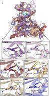
Crystal structure of CX-4945 bound to the active sites of DYRK1A and GSK3β. (A) The overall fold of DYRK1A (violet purple) in cartoon representation with CX-4945 (green sticks) at the ATP-binding pocket. (B) The insert showing CX-4945-DYRK1A interaction in the ATP-binding pocket. (C) Hydrophobic interactions stabilizing CX-4945 at the DYRK1A ATP-binding pocket. (D) Overview of the crystal structure of GSK3β (pink)–CX-4945 (green) complex. (E) Hydrogen-bond interactions of the inhibitor bound to the ATP pocket of GSK3β. (F) Hydrophobic interactions of CX-4945 at the ATP-binding pocket of GSK3β.
Figure 3
Figure 3
Activation loop comparison between phosphorylated and non-phosphorylated Tyr216 GSK3β forms. (A) Superimposition of phosphorylated (pTyr216, pink) and non-phosphorylated (Tyr216, teal) GSK3β with the CX-4945 inhibitor. The activation loop Asp200-Glu226 is shown in yellow. The stabilization of the activation loop in phosphorylated GSK3β (B) and non-phosphorylated GSK3β (C) form.
Figure 4
Figure 4
Representative fluorescence images of CX-4945’s impact on DYRK1A- and GSK3β-mediated inhibition on NFAT signaling. EGFP-NFATc1 (green) was cotransfected with either a mock vector, DYRK1A (red), or GSK3β (red) into HEK293T cells. Cells pretreated for 3 h with CX-4945 or harmine (5 μM) or DMSO before stimulation for 1 h with ionomycin (IM; 6 μM).
Figure 5
Figure 5
Binding mode of CX-4945 in ATP-binding pockets of CMGC and CAMK kinases. (A) Overlay of the crystal structures of kinases from CMGC and CAMK families crystalized with CX-4945. CX-4945 bound to DYRK1A (PDB ID 7Z5N, purple), GSK3β (PDB ID 7Z1F, pink), HIPK2 (PDB ID 6P5S, ruby), CK2α (PDB ID 3PE1, orange), CLK1 (PDB ID 6KHD, wheat), and PIM1 (PDB ID 5O11, slate). (B) Closeup of the binding mode of CX-4945 to the indicated kinases.
Figure 6
Figure 6
Energy decomposition analysis of the complexes between CX-4945 and GSK3β and DYRK1A and CK2α. (A) Maps for electrostatics; (B) lipophilicity maps based on dispersion interactions; (C) total interaction maps; and (D) the magnitude of different contributions to the energy decomposition. For each subfigure, the leftmost part relates to GSK3β, and in the middle, we have DYRK1A and then CK2α. Modeling was based on the PDB files 7Z1G (GSK3β), 7Z5N (DYRK1A), and 3PE1 (CK2α).
Figure 7
Figure 7
Interactions between His160 and CX-4945 in CK2α, as available from the PDB file 3PE1. Protein carbon atoms are represented in light blue, whereas the inhibitor’s carbon atoms are colored in light brown. Hydrogen bonds are marked in green dashed lines. We selected the anchoring of CX-4945’s carboxylate to Lys68 and the water-mediated hydrogen bond with His160. The orange dashed line marks the T-stack interaction.

References

    1. Manning G.; Whyte D. B.; Martinez R.; Hunter T.; Sudarsanam S. The protein kinase complement of the human genome. Science 2002, 298, 1912.10.1126/SCIENCE.1075762. - DOI - PubMed
    1. Buljan M.; Ciuffa R.; van Drogen A.; Vichalkovski A.; Mehnert M.; Rosenberger G.; Lee S.; Varjosalo M.; Pernas L. E.; Spegg V.; Snijder B.; Aebersold R.; Gstaiger M. Kinase Interaction Network Expands Functional and Disease Roles of Human Kinases. Mol. Cell 2020, 79, 504.10.1016/J.MOLCEL.2020.07.001. - DOI - PMC - PubMed
    1. Lahiry P.; Torkamani A.; Schork N. J.; Hegele R. A. Kinase mutations in human disease: interpreting genotype–phenotype relationships. Nat. Rev. Genet. 2010, 11, 60–74. 10.1038/nrg2707. - DOI - PubMed
    1. Cohen P.; Cross D.; Jänne P. A. Kinase drug discovery 20 years after imatinib: progress and future directions. Nat. Rev. Drug Discovery 2021, 20, 551.10.1038/s41573-021-00195-4. - DOI - PMC - PubMed
    1. Attwood M. M.; Fabbro D.; Sokolov A. V.; Knapp S.; Schiöth H. B. Trends in kinase drug discovery: targets, indications and inhibitor design. Nat. Rev. Drug Discovery 2021, 20, 839.10.1038/s41573-021-00252-y. - DOI - PubMed
    2. Blue Ridge Institute for Medical Research. https://brimr.org/protein-kinase-inhibitors/ (accessed Dec 28, 2022).

Publication types

MeSH terms