

Tracheal Mirror Image Artifact in Patients With Normal and Pathologic Tracheas
- PMID: 36883989
- PMCID: PMC10485171
- DOI: 10.1002/ohn.316
Tracheal Mirror Image Artifact in Patients With Normal and Pathologic Tracheas
Abstract
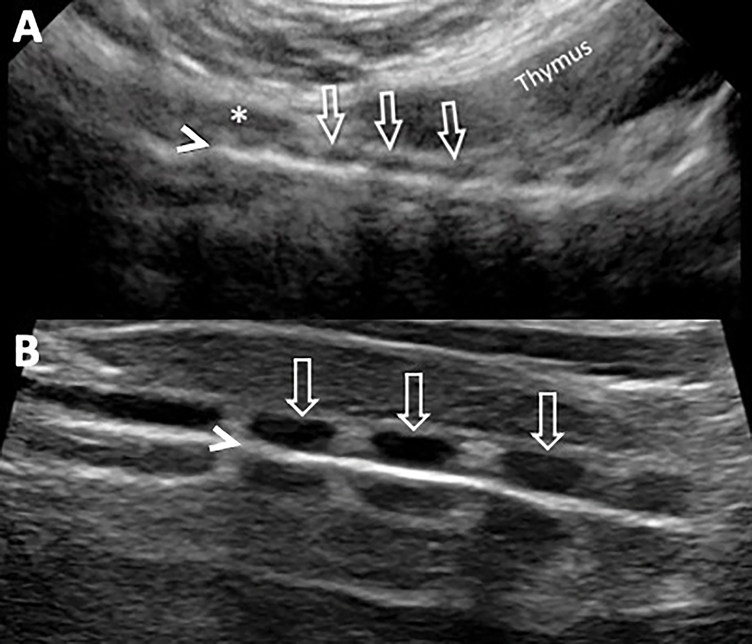
Ultrasonography is gaining popularity as a diagnostic imaging modality for airway pathology. Tracheal ultrasound (US) has several nuances that are important for clinicians, including imaging artifacts, which can be mistaken for pathology. Tracheal mirror image artifacts (TMIAs) occur when the US beam is reflected back to the transducer in a nonliner direction or with multiple timesteps. It has previously been believed that the convexity of the tracheal cartilage prevents mirror image artifacts, but in reality, the air column acts as an acoustic mirror and causes TMIA. We describe a cohort of patients with both normal and pathologic tracheas, all of whom have TMIA on the tracheal US. These artifacts are important to recognize, especially as the airway US becomes more commonplace.
Keywords: airway; artifact; mirror image; trachea; tracheal cartilaginous sleeve; ultrasound.
© 2023 American Academy of Otolaryngology-Head and Neck Surgery Foundation.
Conflict of interest statement
Conflicts of interest: The authors declare that there are no conflicts of interest
Figures

References
-
- You-Ten KE, Siddiqui N, Teoh WH, et al. Point-of-care ultrasound (Pocus) of the upper airway. Can J Anesth/J Can Anesth. 2018;65(4):473–484. - PubMed
-
- Liao LJ, Wen MH, Yang TL. Point-of-care ultrasound in otolaryngology and head and neck surgery: A prospective survey study. J Formos Med Assoc. 2021;120(8):1547–1553. - PubMed
-
- Feldman MK, Katyal S, Blackwood MS. US artifacts. Radiographics. 2009;29(4):1179–1189. - PubMed
-
- Kerr DM, Middleton WD. Reflections on the ultrasound mirror image artifact. Ultrasound Q. 2020;36(4):287–299. - PubMed
-
- Diazzi C, Gnarini V, Brigante G, et al. Ultrasound mirror artifact of a thyroid nodule by trachea mimicking a tracheal mass. Thyroid. 2011;21(8):929–930. - PubMed
Publication types
MeSH terms
Grants and funding
LinkOut - more resources
Full Text Sources
