. 2023 Mar 31;132(7):867-881.
doi: 10.1161/CIRCRESAHA.122.321583. Epub 2023 Mar 8.

β3AR-Dependent Brain-Derived Neurotrophic Factor (BDNF) Generation Limits Chronic Postischemic Heart Failure

Affiliations

β3AR-Dependent Brain-Derived Neurotrophic Factor (BDNF) Generation Limits Chronic Postischemic Heart Failure

Alessandro Cannavo et al. Circ Res. .

Abstract

Background: Loss of brain-derived neurotrophic factor (BDNF)/TrkB (tropomyosin kinase receptor B) signaling accounts for brain and cardiac disorders. In neurons, β-adrenergic receptor stimulation enhances local BDNF expression. It is unclear if this occurs in a pathophysiological relevant manner in the heart, especially in the β-adrenergic receptor-desensitized postischemic myocardium. Nor is it fully understood whether and how TrkB agonists counter chronic postischemic left ventricle (LV) decompensation, a significant unmet clinical milestone.

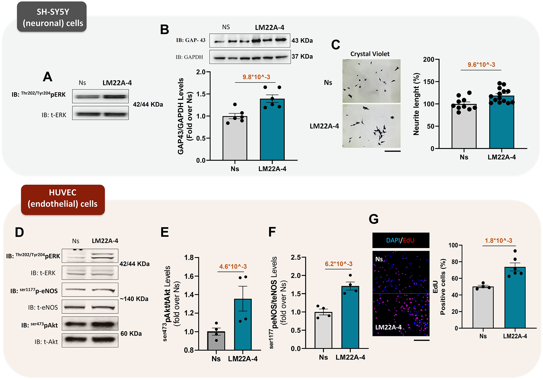
Methods: We conducted in vitro studies using neonatal rat and adult murine cardiomyocytes, SH-SY5Y neuronal cells, and umbilical vein endothelial cells. We assessed myocardial ischemia (MI) impact in wild type, β3AR knockout, or myocyte-selective BDNF knockout (myoBDNF KO) mice in vivo (via coronary ligation [MI]) or in isolated hearts with global ischemia-reperfusion (I/R).

Results: In wild type hearts, BDNF levels rose early after MI (<24 hours), plummeting at 4 weeks when LV dysfunction, adrenergic denervation, and impaired angiogenesis ensued. The TrkB agonist, LM22A-4, countered all these adverse effects. Compared with wild type, isolated myoBDNF KO hearts displayed worse infarct size/LV dysfunction after I/R injury and modest benefits from LM22A-4. In vitro, LM22A-4 promoted neurite outgrowth and neovascularization, boosting myocyte function, effects reproduced by 7,8-dihydroxyflavone, a chemically unrelated TrkB agonist. Superfusing myocytes with the β3AR-agonist, BRL-37344, increased myocyte BDNF content, while β3AR signaling underscored BDNF generation/protection in post-MI hearts. Accordingly, the β1AR blocker, metoprolol, via upregulated β3ARs, improved chronic post-MI LV dysfunction, enriching the myocardium with BDNF. Last, BRL-37344-imparted benefits were nearly abolished in isolated I/R injured myoBDNF KO hearts.

Conclusions: BDNF loss underscores chronic postischemic heart failure. TrkB agonists can improve ischemic LV dysfunction via replenished myocardial BDNF content. Direct cardiac β3AR stimulation, or β-blockers (via upregulated β3AR), is another BDNF-based means to fend off chronic postischemic heart failure.

Keywords: brain-derived neurotrophic factor; cardiac disorders; heart failure; myocardial ischemia; receptors, adrenergic.


Figures

Figure 1.
Figure 1.. Post-ischemic LV dysfunction is coupled with lower cardiac BDNF content and associated with reduced cardiac sympathetic nerve fibers and poor vascularization.
A) Mouse genotype: WT; Intervention: MI; B-C) Representative immunoblots/densitometric quantitative analysis of multiple independent experiments to evaluate BDNF expression levels, in total cardiac lysates of 6 hours (hrs) and 24 hrs (sham n=3, 6 hrs n=3, and 24 hrs n=4) (B) or 1 week and 4 weeks (sham n=4, 1wk n=5, and 4wks n=4) (C) post-MI mice. Sham-operated animals were used as control. GAPDH levels were used as loading control. D) Representative panels (merge) of DAPI (blue), α-sarcomeric actinin staining (green), and BDNF (red) immunofluorescence images (scale bar, 50 μm) showing data concerning BDNF expression in cardiac sections from sham, 6 hrs, 24 hrs, 1 and 4 weeks post-MI mice; E-F-G) Representative images of echocardiographic analysis (M-mode) performed at 4 weeks post-MI and dot plots showing F) left ventricle (LV) ejection fraction (EF, %) (sham n=5, and MI n=4), G) LV internal diameter at diastole (LVIDd, mm) (sham n=5, and MI n=4) of individual mice from each of the groups: sham and MI. H) Representative images/aquantitative data of percent cardiac fibrosis (Picro-Sirius red staining, Scale bar 200 μm) in cardiac sections from sham and MI mice (sham n=5, and MI n=4). I) Representative images of Lectin Bandeiraea simplicifolia I staining of capillaries in the ischemic vs. sham-operated myocardium (scale bar: 200 μm, left panels); and a bar graph showing capillary/mm2 in cardiac sections of sham and MI mice (sham n=3, MI remote n=3, and MI border n=3). J) Representative images/quantitative data of percent tyrosine hydroxylase (TH) positive (+) fibers (immunofluorescence staining, scale bar (50 μm) in cardiac sections from sham and MI mice (sham n=5, and MI n=4). K) Representative immunoblots/densitometric quantitative analysis of TH protein level in total cardiac lysates of sham and MI mice (sham n=4, and MI n=4). GAPDH levels were used as a loading control. Data were analyzed utilizing a nonparametric rank-based test with Shaffer post hoc correction (B, C, I, F, G, H, J, and K). All data are shown as mean±s.e.m.
Figure 2.
Figure 2.. TrkB-agonism via LM22A-4 enhances neuronal and endothelial cell function, in vitro.
A) Representative immunoblots showing ERK activation (phospho-Thr202/Tyr204) in total lysates from SH-SY5Y neuronal cell lysates unstimulated (Ns) or stimulated with LM22A-4 (100 nM) for 10 min. Total ERK (tERK) levels were used as loading control. B) Representative immunoblots/densitometric quantitative analysis of GAP-43 levels in total lysates from SH-SH5Y neuronal cell lysates Ns or stimulated with LM22A-4 (100 nM) for 24 hours (Ns n=6, and LM22A-4 n=6). GAPDH levels were used as loading control. C) Representative images/quantitative data showing neurite length percentage (%) in SH-SY5Y Ns or stimulated with LM22A-4 (100 nM) for 24 hours (Ns n=10, LM22A-4 n=14). D-E-F) Representative immunoblots (D) and densitometric quantitative analysis (E-F) showing ERK activation (phospho-Thr202/Tyr204) (D); Akt activation (phospho-ser473) (E) and eNOS activation (phospho-ser1177) levels (F) in total lysates from HUVEC lysates Ns or stimulated with LM22A-4 (100 nM) for 10 min (Ns n=4, and LM22A-4 n=4). Total ERK (tERK), tAkt, and eNOS levels were used as the loading control, respectively. G) Representative images/quantitative data of the EdU positive cell percentage (%) (immunofluorescence staining, scale bar (200 μm) in HUVEC cells. Ns or stimulated with LM22A-4 (100 nM) for 12 hours (Ns n=4, and LM22A-4 n=6). Data were analyzed via a nonparametric rank-based test with Shaffer post hoc correction (B, E, F, G, and C). All data are shown as mean±s.e.m.
Figure 3.
Figure 3.. LM22A-4 and 7,8-DHF enhance inotropy and whole calcium transient in isolated adult murine cardiomyocyte via TrkB activation.
A-C) Representative image of adult murine isolated cardiomyocyte (scale bar 25 μm) (A) and quantitative data showing percentage change of sarcomere shortening (B) and percentage change of calcium transient (C) of isolated cardiomyocytes unstimulated (0 μM; n=10) or stimulated with LM22A-4 at 2.5 μM (n=10), 5 μM (n=10), and 10 μM (B, n=9 and C, n=10) D-E) Quantitative data showing percentage change of sarcomere shortening (D) and percentage change of calcium transient (E) of isolated cardiomyocytes unstimulated (0 μM) or stimulated with 7,8-DHF at 2.5 μM, 5 μM, and 10 μM (0 μM n= 8, 2.5 μM n=8, 5 μM n=8, and 10 μM n=8). F) Representative immunoblots and densitometric quantitative analysis showing ERK activation (phospho-Thr202/Tyr204) in total lysates from NRVMs Ns or stimulated with LM22A-4 (100 nM) or 7,8-DHF (100 nM) for 10 min (Ns n=8, LM22A-4 n=6, and 7,8-DHF n=6). Total ERK (tERK), levels were used as the loading control. G-H) Representative immunoblots and densitometric quantitative analysis showing (G) ERK activation (phospho-Thr202/Tyr204) and (H) TrkB phosphorylation (phospho-Tyr816) levels in total lysates from NRVMs Ns or stimulated with LM22A-4 (100 nM) for 10 min. Prior LM22A-4 stimulation a group of cells was pre-treated with ANA-12 (10 μM) for 30 min. (Ns n=5, LM22A-4 n=5, and A12/LM22A-4 n=5). Total ERK (tERK), levels were used as the loading control. Data were analyzed employing a nonparametric rank-based test with Shaffer post hoc correction (B, D,C, E, F, G, and H). All data are shown as mean±s.e.m.
Figure 4.
Figure 4.. In vivo TrkB-agonism prevents chronic post-ischemic cardiac decompensation and increases myocardial BDNF content.
A) Mouse genotype: WT; Interventions: MI ± LM22A-4; B) Dot plots showing the percentage of infarct size evaluated 4 weeks post-MI in mice treated with vehicle (saline solution, MI) or LM22A-4 (MI n=6, and MI+LM22A-4 n=8). C-D-E) Dot plots showing the echocardiographic analysis performed at 4 weeks post-MI (C) LV ejection fraction (EF, %), (D) LV internal diameter at diastole (LVIDd, mm), (E) LV internal diameter at systole (LVIDs, mm) (sham n=12, MI n=8, and MI+LM22A-4 n=8). F) Representative images/quantitative data showing the percentage of cardiac fibrosis (Picro-Sirius red staining, scale bar 200 μm) in cardiac sections from sham, MI, and MI+LM22A4 mice (sham n=7, MI n=6, and MI+LM22A-4 n=6). G) Representative images of Lectin Bandeiraea simplicifolia I staining of capillaries in the ischemic vs. sham-operated myocardium (scale bar: 200 μm) and a bar graph showing capillary/mm2 in cardiac section of sham, MI and MI+LM22A-4 mice (sham n=6, MI remote n=4, MI border n=4, MI+LM22A-4 remote n=5, and MI+LM22A4 n=4). H) Representative images/quantitative data of TH+ fiber percentage (immunofluorescence staining, scale bar (50 μm) in cardiac sections from sham, MI and MI+LM22A-4 mice (sham n=3, MI n=3, and MI+LM22A4 n=4). I) Representative immunoblots/densitometric quantitative analysis showing of BDNF levels in total cardiac lysates of MI and MI+LM22A4 mice (sham n=4, MI n=4, and MI+LM22A-4 n=4). GAPDH levels were used as a loading control. Data were analyzed using a nonparametric rank-based test with Shaffer post hoc correction (B, C, D, E, F, G, H, and I). All data are shown as mean±s.e.m.
Figure 5.
Figure 5.. Ex Vivo TrkB agonism limits infarct size and cardiac functional deterioration post-ischemia-reperfusion injury.
A) Mouse genotypes: WT and myoBDNF KO; Interventions: I/R injury± LM22A-4; B-C) Representative images of I/R induced infarct size (B) and quantitative data of the infarct size (C) by global ischemia via Langendorff perfusion with or without LM22A-4 (20 μM) during the first 10 minutes of reperfusion. (WT n=5, WT+LM22A-4 n=9, myoBDNF KO n=5, and myoBDNF KO+LM22A-4 n=7). D) Quantitative data showing cTnI release in the coronary effluent. (WT Baseline n=5, WT 2 hrs reperfusion n=5, myoBDNF KO Baseline n=5, and myoBDNF KO 2 hrs reperfusion n=5). (E), heart rate (F), LV developed pressure (G), dP/dt max (H), and dP/dtmin (I) post I/R injury via Langendorff perfusion with or without LM22A-4 (20 μM) during the first 10 minutes of reperfusion. (WT n=7, WT+LM22A-4 n=9, myoBDNF KO n=7, and myoBDNF KO+LM22A-4 n=7). J) Representative images of TUNEL assay and quantitative data showing the percentage of TUNEL positive cells (%) (immunofluorescence staining, scale bar (50 μm) in I/R induced hearts of WT and myoBDNF KO. (WT n=8, and myoBDNF KO n=8). Data were analyzed utilizing a nonparametric rank-based test with Shaffer post hoc correction (C, D, E, G, H, I, and J). All data are shown as mean±s.e.m.
Figure 6.
Figure 6.. In ischemic CHF, disrupted β3AR-signaling accounts for BDNF expression loss, reduced cardiac sympathetic innervation, and angiogenesis.
A) Representative immunoblots and densitometric quantitative analysis showing levels of BDNF in total lysates from NRVMs unstimulated (Ns) or stimulated with NEpi (10 μM) or BRL-37344 (BRL, 1 μM) for 12 hrs. GAPDH was the loading control. (Ns n=3, Nepi n=3, and BRL n=3). B) Representative immunoblots/densitometric quantitative analysis of BDNF levels in total lysates from unstimulated (Ns) or NEpi-stimulated (10 μM for 12 hrs) NRVMs. Prior NEpi treatment, a group of cells was pre-treated with SR59230A (SR, 10 μM) for 30 min. GAPDH was the loading control. (Ns n=6, Nepi n=5, SR n=4, and SR/Nepi n=6). C) Representative immunoblots/densitometric quantitative analysis of BDNF levels in total lysates from NRVMs unstimulated (Ns) or stimulated with BRL (1 μM) for 12 hrs. Prior BRL treatment a group of cells were pre-treated with SR59230A (SR, 10 μM) for 30 min. GAPDH was used as loading control. (Ns n=4, BRL n=4, and SR/BRL n=4). D) Mouse genotypes: WT and β3AR KO mice; Intervention: MI. E-F-G) Representative immunoblots (E) and densitometric quantitative analysis (F-G) showing BDNF and TH levels in total cardiac lysates from the following groups: WT (sham and MI) and β3AR KO (sham and MI) mice. GAPDH levels were used for protein loading controls. F) BDNF: WT sham n=5, WT MI sham n=6, β3AR KO sham n=5, and β3AR KO MI n=6 and G) TH: WT sham n=7, WT MI sham n=8, β3AR KO sham n=7, and β3AR KO MI n=8. H) Representative images/quantitative data of TH+ fiber percentage (immunofluorescence staining, scale bar (50 μm) in cardiac sections from sham, MI (WT vs. β3AR KO) mice (WT sham n=4, β3AR KO sham n=4, and β3AR KO MI n=4). I) Representative images of Lectin Bandeiraea simplicifolia I staining of capillaries in the ischemic vs. sham-operated myocardium (scale bar: 200 μm) and bar graph showing capillary/mm2 in cardiac section from sham and MI (WT vs. β3AR KO) mice (WT remote sham n=8, β3AR KO remote sham n=6, WT remote MI n=7, β3AR KO remote MI n=5, WT border MI n=7, and β3AR KO border MI n=5). Data were analyzed using a nonparametric rank-based test with Shaffer post hoc correction (A, B, C, F, G, H, and I). All data are shown as mean±s.e.m.
Figure 7.
Figure 7.. In I/R injured hearts, β3AR benefits require cardiomyocyte borne BDNF.
A) Mouse genotypes: WT and myoBDNF KO; Interventions: I/R injury ±BRL-37344. B-C) Representative images of I/R induced infarct size (TTC staining) (B) and quantitative data of the infarct size (C) by global ischemia via Langendorff perfusion with or without BRL37344 (10 μM) during first 10 minutes of reperfusion (WT n=5, WT+BRL37344 n=10, myoBDNF KO n=5, and myoBDNF KO+BRL37344 n=7). D-H) Quantitative data showing percentage recovery of rate-pressure product (D), heart rate (E), LV developed pressure (F), dP/dtmax (G) and dP/dtmin (H). (WT n=7, WT+BRL37344 n=9, myoBDNF n=7, and myoBDNF+BRL37344 n=7). (I) Representative images/quantitative data of TUNEL positive cell percentage (%) (immunofluorescence staining, scale bar (50 μm) in I/R induced hearts of WT and myoBDNF KO with BRL37344. (WT+BRL37344 n=9, and myoBDNF KO+BRL37344 n=9). Data were analyzed via a nonparametric rank-based test with Shaffer post hoc (C, D, GF, H, and I). All data are shown as mean±s.e.m.
Figure 8.
Figure 8.. Synopsis of the findings/Conceptual framework of the study.
TrkB agonism and β3AR-induced BDNF production arrest post-ischemic CHF progression, exerting myocardial autocrine/paracrine protective effects: 1) inducing a therapeutic response in ischemic cardiomyocytes, i.e., limiting cell death and improving function; 2) activating endothelial cell proliferation, and 3) enhancing autonomic neuronal sprouting.

References

    1. Johansson S, Rosengren A, Young K, Jennings E. Mortality and morbidity trends after the first year in survivors of acute myocardial infarction: a systematic review. BMC Cardiovasc Disord. 2017;17:53. doi: 10.1186/s12872-017-0482-9 - DOI - PMC - PubMed
    1. Benarroch EE. Brain-derived neurotrophic factor: Regulation, effects, and potential clinical relevance. Neurology. 2015;84:1693–1704. doi: 10.1212/WNL.0000000000001507 - DOI - PubMed
    1. Kermani P, Hempstead B. BDNF Actions in the Cardiovascular System: Roles in Development, Adulthood and Response to Injury. Front Physiol. 2019;10:455. doi: 10.3389/fphys.2019.00455 - DOI - PMC - PubMed
    1. Feng N, Huke S, Zhu G, Tocchetti CG, Shi S, Aiba T, Kaludercic N, Hoover DB, Beck SE, Mankowski JL, et al. Constitutive BDNF/TrkB signaling is required for normal cardiac contraction and relaxation. Proc Natl Acad Sci U S A. 2015;112:1880–1885. doi: 10.1073/pnas.1417949112 - DOI - PMC - PubMed
    1. Fulgenzi G, Tomassoni-Ardori F, Babini L, Becker J, Barrick C, Puverel S, Tessarollo L. BDNF modulates heart contraction force and long-term homeostasis through truncated TrkB.T1 receptor activation. J Cell Biol. 2015;210:1003–1012. doi: 10.1083/jcb.201502100 - DOI - PMC - PubMed

Publication types

Substances

LinkOut - more resources