

Clinical identification and endodontic management of furcation canals: a case series
- PMID: 36888840
- PMCID: PMC10027094
- DOI: 10.1590/0103-6440202304817
Clinical identification and endodontic management of furcation canals: a case series
Abstract
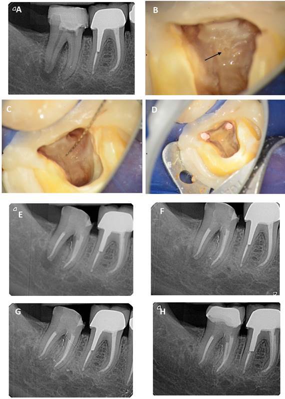
In the case of endodontic infection, the presence of furcation canals can be at the origin of a periodontal lesion located in the furcation. The furcation being very close to the marginal periodontium, this type of lesion can be favorable to the genesis of an endo-periodontal lesion. These furcation canals are lateral canals located on the pulp chamber floor and constitute one of the many physiological communication pathways between endodontic and periodontal tissues. These canals are most often difficult to localize, shape, and to fill because of their small diameter and length. The disinfection of the pulp chamber floor with sodium hypochlorite solution may contribute to the disinfection of furcation canals when they are not identified, shaped, and/or filled. This case series illustrates the endodontic management of visible furcation canals associated with an endo-periodontal lesion. These furcation canals had a large diameter which allowed their identification during the endodontic treatment.
Figures

References
-
- Tavares PB, Bonte E, Boukpessi T, Siqueira JF, Jr, Lasfargues JJ. Prevalence of apical periodontitis in root canal-treated teeth from an urban French population: influence of the quality of root canal fillings and coronal restorations. J Endod. 2009;35(6):810–813. - PubMed
-
- Wolcott J, Ishley D, Kennedy W, Johnson S, Minnich S. Clinical investigation of second mesiobuccal canals in endodontically treated and retreated maxillary molars. J Endod. 2002;28(6):477–479. - PubMed
-
- Seltzer S, Bender IB, Ziontz M. The interrelationship of pulp and periodontal disease. Oral Surgery, Oral Medicine, Oral Pathology. 1963;16(12):1474–1490. - PubMed
-
- Barrett M. The internal anatomy of the teeth with special reference to the pulp with its branches. Dental Cosmos. 1925;67:581–592.
-
- Rotstein I, Simon JHS. Diagnosis, prognosis and decision-making in the treatment of combined periodontal-endodontic lesions. Periodontol. 2004;34(1):165–203. 2000. - PubMed
MeSH terms
LinkOut - more resources
Full Text Sources
