. 2023 Mar 8;13(1):3868.
doi: 10.1038/s41598-023-29915-z.

A screen for MeCP2-TBL1 interaction inhibitors using a luminescence-based assay

Affiliations

A screen for MeCP2-TBL1 interaction inhibitors using a luminescence-based assay

Beatrice Alexander-Howden et al. Sci Rep. .

Abstract

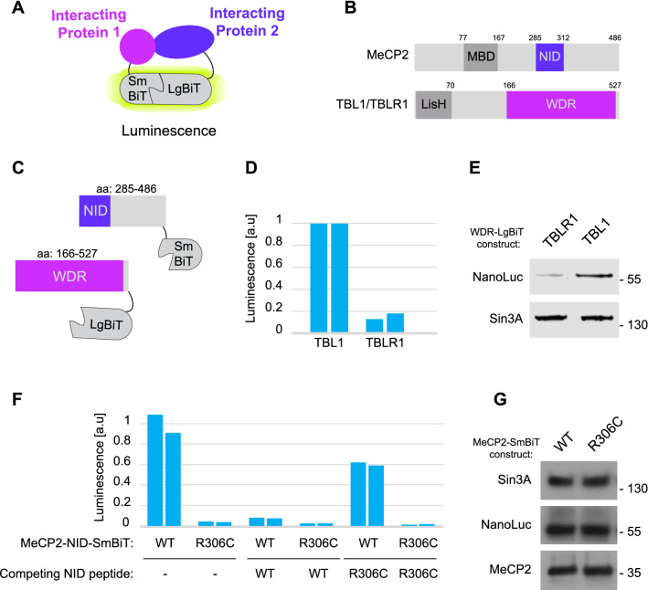
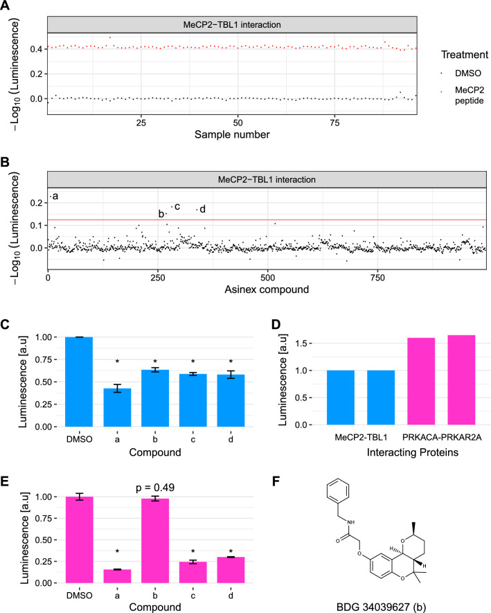
Understanding the molecular pathology of neurodevelopmental disorders should aid the development of therapies for these conditions. In MeCP2 duplication syndrome (MDS)-a severe autism spectrum disorder-neuronal dysfunction is caused by increased levels of MeCP2. MeCP2 is a nuclear protein that binds to methylated DNA and recruits the nuclear co-repressor (NCoR) complex to chromatin via an interaction with the WD repeat-containing proteins TBL1 and TBLR1. The peptide motif in MeCP2 that binds to TBL1/TBLR1 is essential for the toxicity of excess MeCP2 in animal models of MDS, suggesting that small molecules capable of disrupting this interaction might be useful therapeutically. To facilitate the search for such compounds, we devised a simple and scalable NanoLuc luciferase complementation assay for measuring the interaction of MeCP2 with TBL1/TBLR1. The assay allowed excellent separation between positive and negative controls, and had low signal variance (Z-factor = 0.85). We interrogated compound libraries using this assay in combination with a counter-screen based on luciferase complementation by the two subunits of protein kinase A (PKA). Using this dual screening approach, we identified candidate inhibitors of the interaction between MeCP2 and TBL1/TBLR1. This work demonstrates the feasibility of future screens of large compound collections, which we anticipate will enable the development of small molecule therapeutics to ameliorate MDS.


Conflict of interest statement

The authors declare no competing interests.

Figures

Figure 1
Figure 1
NanoLuc based protein complementation assay for MeCP2-TBL1 binding. (A) Schematic showing interaction between two proteins of interest bridging the large (LgBiT) and small (SmBiT) fragments of NanoLuc resulting in luminescence. (B) Functional domains of MeCP2 and TBL1/TBLR1. The MBD (methyl-CpG binding domain) is the DNA binding domain of MeCP2. The NID of MeCP2 mediates binding to the NCoR complex via an interaction with TBL1/TBLR1. The WD repeat (WDR) domain of TBL1/TBLR1 binds to the NID of MeCP2. The Lis homology (LisH) domain of TBL1/TBLR1 mediates self-association and incorporation into the NCoR complex. (C) Illustration of the MeCP2-SmBiT and TBL1/TBLR1-LgBiT fusion proteins used in this study. SmBiT is fused to a C-terminal fragment of MeCP2 (residues 285–486). LgBiT is fused to the WD repeat (WDR) domain of TBL1/TBLR1 (residues 166–527 for TBL1). (D) Robust luminescence is observed using extracts from cells transiently expressing MeCP2-SmBiT and TBL1/TBLR1-LgBiT. The two bars represent the results of biological replicates (n = 2) for each condition. (E) Western blotting with antibodies against NanoLuc and Sin3A (loading control) reveals stronger expression of the TBL1-LgBiT fusion protein than of TBLR1-LgBiT. (F) Luminescence observed with extracts from cells co-expressing MeCP2-SmBiT and TBL1-LgBiT is strongly reduced by introducing the R306C mutation into the NID of MeCP2. Luminescence is also inhibited by the addition of 20 µM of a peptide corresponding to the NID of MeCP2. A similar peptide carrying the R306C mutation has a much reduced inhibitory effect. The two bars represent the results of biological replicates (n = 2) for each condition. (G) Western blotting for MeCP2, NanoLuc and Sin3A (loading control) shows that the R306C mutation in MeCP2 does not affect the expression levels of the MeCP2-SmBiT and TBL1-LgBiT fusion proteins.
Figure 2
Figure 2
Screening for MeCP2-TBL1 inhibitors in a protein–protein interaction inhibitor library. (A) Z-factor determination using a short sequence-optimized MeCP2 peptide (MeCP2 K304Y, residues 298–309 of MeCP2) (red) at 50 μM as a positive control and DMSO (black) as a negative control. The measured luminescence for each sample was normalized to the mean luminescence observed with DMSO. The y-axis shows the -log10 of these normalized values. (B) Distribution of hits from a screen of 995 compounds from a custom Asinex protein–protein interaction inhibitor library assayed at 20 µM. The interaction between MeCP2 and TBL1 was monitored via the luminescence signal in a NanoLuc complementation assay. Measured luminescence for each compound was normalized to the mean luminescence value obtained with DMSO. The y-axis shows the − log10 of these normalized values. Four compounds (labelled a–d) show greater than 25% inhibition (threshold indicated by the red line). (C) Individual validation of the top four hits (n = 3; * p < 0.005 (t-test)). Error bars represent standard deviations. For all four compounds the degree of inhibition achieved at 20 µM was consistent with that observed in the original screen. (D) Robust luminescence observed using extracts from cells transiently expressing NanoLuc fragments as fusion proteins with two subunits of protein kinase A (SmBiT-PRKACA and LgBiT-PRKAR2A). The signal was approximately 1.6-fold greater than is observed using the MeCP2-TBL1 interaction assay. The two bars represent the results of biological replicates (n = 2) for each condition. (E) Specificity of primary Asinex library hits in the PKA control assay (n = 3; * p < 0.005 (t-test)). Error bars represent standard deviations. Only compound BDG 34039627 (b) did not affect luminescence in the PKA control assay. (F) Chemical structure of BDG 34039627.
Figure 3
Figure 3
Screening of an FDA-approved drug library with the MeCP2-TBL1 assay and PRKACA-PRKAR2A counter assay. (A) Distribution of hits from a screen of 1981 compounds from the ApexBio FDA-approved Drug Library assayed at 20 µM. The interaction between MeCP2 and TBL1 was monitored via the luminescence signal in a NanoLuc complementation assay (top panel). Also shown is the distribution of hits from a control screen using a NanoLuc complementation assay based on the interaction between PRKACA and PRKAR2A (middle panel). For the top and middle panels the measured luminescence for each compound was normalized to the mean luminescence obtained with DMSO. The y-axis shows the − log10 of these normalized values. The bottom panel shows inhibition in the MeCP2-TBL1 assay after adjusting for activity of compounds in the PRKACA-PRKAR2A assay. The top five hits (e–i) are indicated above the 35% inhibition threshold (red line). (B) Chemical structures of the non-peptide hits from dual screening of the ApexBio library with the MeCP2-TBL1 assay and the control PRKACA-PRKAR2A assay. (C) Individual validation of the top five hits from the ApexBio library screen against both the MeCP2-TBL1 and the PRKACA-PRKAR2A NanoLuc complementation assays. At 20 µM, flunarizine, luminespib, oridonin, and the alpha-1 antitrypsin fragment 235–243 all achieved statistically significantly greater inhibition in the MeCP2-TBL1 assay than in the control assay (n = 7; * p < 0.001 (t-test)). Error bars represent standard deviations. BIX02189 did not have a statistically different effect between the two assays (n = 7; p = 0.47 (t-test)).

References

    1. Amir RE, Van den Veyver IB, Wan M, Tran CQ, Francke U, Zoghbi HY. Rett syndrome is caused by mutations in X-linked MECP2, encoding methyl-CpG-binding protein 2. Nat. Genet. 1999;23:185–188. doi: 10.1038/13810. - DOI - PubMed
    1. Percy AK, Neul JL, Glaze DG, Motil KJ, Skinner SA, Khwaja O, Lee H-S, Lane JB, Barrish JO, Annese F, et al. Rett syndrome diagnostic criteria: Lessons from the Natural History Study. Ann. Neurol. 2010;68:951–955. doi: 10.1002/ana.22154. - DOI - PMC - PubMed
    1. Neul JL, Kaufmann WE, Glaze DG, Christodoulou J, Clarke AJ, Bahi-Buisson N, Leonard H, Bailey MES, Schanen NC, Zappella M, et al. Rett syndrome: Revised diagnostic criteria and nomenclature. Ann. Neurol. 2010;68:944–950. doi: 10.1002/ana.22124. - DOI - PMC - PubMed
    1. Rett A. On a unusual brain atrophy syndrome in hyperammonemia in childhood. Wien Med. Wochenschr. 1966;116:723–726. - PubMed
    1. Hagberg B, Aicardi J, Dias K, Ramos O. A progressive syndrome of autism, dementia, ataxia, and loss of purposeful hand use in girls: Rett’s syndrome: report of 35 cases. Ann. Neurol. 1983;14:471–479. doi: 10.1002/ana.410140412. - DOI - PubMed

Publication types

MeSH terms

Substances

LinkOut - more resources