

Histological confinement of transglutaminase-mediated nit sheath crosslinking is essential for proper oviposition and egg coating in the human head louse, Pediculus humanus capitis
- PMID: 36890607
- PMCID: PMC9997029
- DOI: 10.1186/s13071-023-05720-5
Histological confinement of transglutaminase-mediated nit sheath crosslinking is essential for proper oviposition and egg coating in the human head louse, Pediculus humanus capitis
Abstract
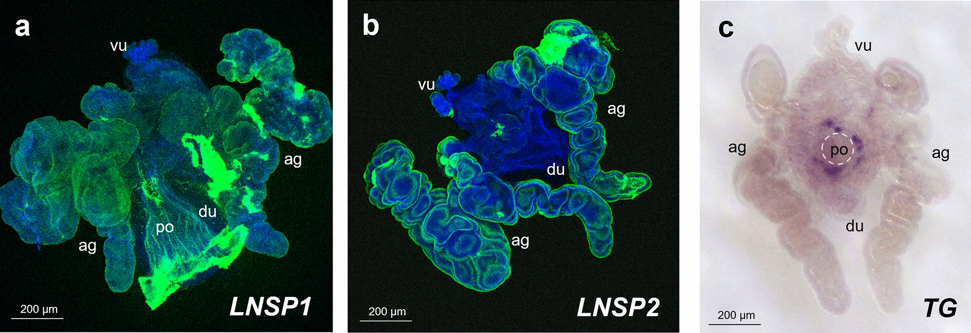
Background: Head louse females secrete liquid gel, which is mainly composed of the louse nit sheath protein 1 (LNSP1) and LNSP2, when they lay eggs. The gel is crosslinked by transglutaminase (TG) to form the nit sheath, which covers most of the egg except the top operculum area where breathing holes are located. Knowledge on the selective mechanism of nit sheath solidification to avoid uncontrolled crosslinking could lead to designing a novel method of louse control, but no information is available yet.
Methods: To elucidate the crosslinking mechanisms of nit sheath gel inside the reproductive system of head louse females, in situ hybridization in conjunction with microscopic observation of the oviposition process was conducted.
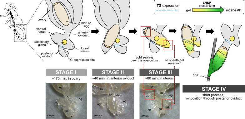
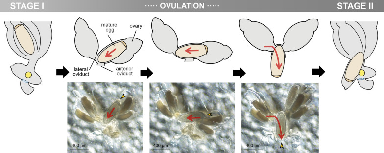
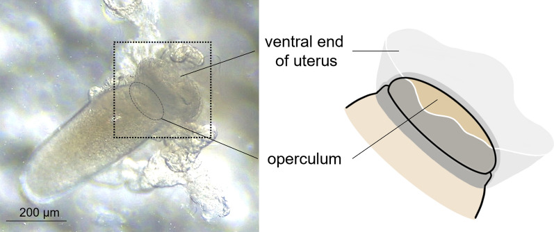
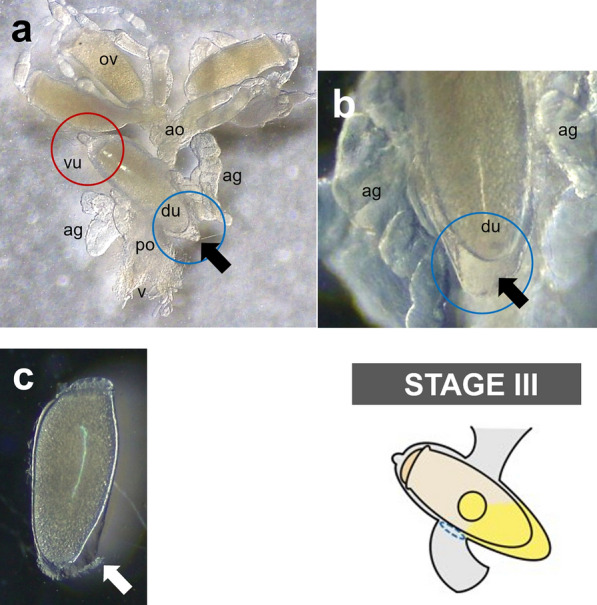
Results: Histochemical analysis revealed that LNSP1 and LNSP2 are expressed over the entire area of the accessory gland and uterus, whereas TG expression site is confined to a highly localized area around the opening of posterior oviduct. Detailed microscopic observations of oviposition process uncovered that a mature egg is positioned in the uterus after ovulation. Once aligned inside the uterus, the mature egg is redirected so that its operculum side is tightly held by the ventral end of the uterus being positioned toward the head again and its pointed bottom end being positioned toward the dorsal end of the uterus, which functions as a reservoir for the nit sheath gel.
Conclusions: Physical separation of the TG-mediated crosslinking site from the ventral end of the uterus is necessary to avoid uncontrolled crosslinking inside the uterus and to ensure selective crosslinking over only the lower part of egg without any unwanted crosslinking over the operculum during oviposition.
Keywords: Egg sheath; Head louse; LNSP; Nit; Oviposition; Pediculus humanus capitis; Transglutaminase.
© 2023. The Author(s).
Conflict of interest statement
The authors declare no competing interests.
Figures
Update of
-
Histological confinement of transglutaminase-mediated nit sheath crosslinking is essential for proper oviposition and egg coating in the human head louse, Pediculus humanus capitis.Res Sq [Preprint]. 2023 Feb 9:rs.3.rs-2559266. doi: 10.21203/rs.3.rs-2559266/v1. Res Sq. 2023. Update in: Parasit Vectors. 2023 Mar 9;16(1):93. doi: 10.1186/s13071-023-05720-5. PMID: 36798255 Free PMC article. Updated. Preprint.
References
-
- Ferris GF. The sucking lice. Mem Pacif Coast Ent Soc. 1951 1.
-
- Nuttall GH. The biology of Pediculus humanus. Parasitology. 1919;11:201–220. doi: 10.1017/S0031182000004194. - DOI
-
- Carter DG. Insect egg glue: an investigation of the nature and secretion of insect egg glues, with special reference to the human louse, Pediculus humanus and the cabbage white butterfly. Pieris: University of Cambridge; 1990.
MeSH terms
Grants and funding
LinkOut - more resources
Full Text Sources
Medical
Research Materials
Miscellaneous
