Review
. 2023 Feb 20:14:1122409.
doi: 10.3389/fimmu.2023.1122409. eCollection 2023.

The mast cell: A Janus in kidney transplants

Affiliations
Review

The mast cell: A Janus in kidney transplants

G van der Elst et al. Front Immunol. .

Erratum in

  • Corrigendum: The mast cell: A Janus in kidney transplants.
    van der Elst G, Varol H, Hermans M, Baan CC, Duong-van Huyen JP, Hesselink DA, Kramann R, Rabant M, Reinders MEJ, von der Thüsen JH, van den Bosch TPP, Clahsen-van Groningen MC. van der Elst G, et al. Front Immunol. 2023 Mar 21;14:1183969. doi: 10.3389/fimmu.2023.1183969. eCollection 2023. Front Immunol. 2023. PMID: 37026016 Free PMC article.

Abstract

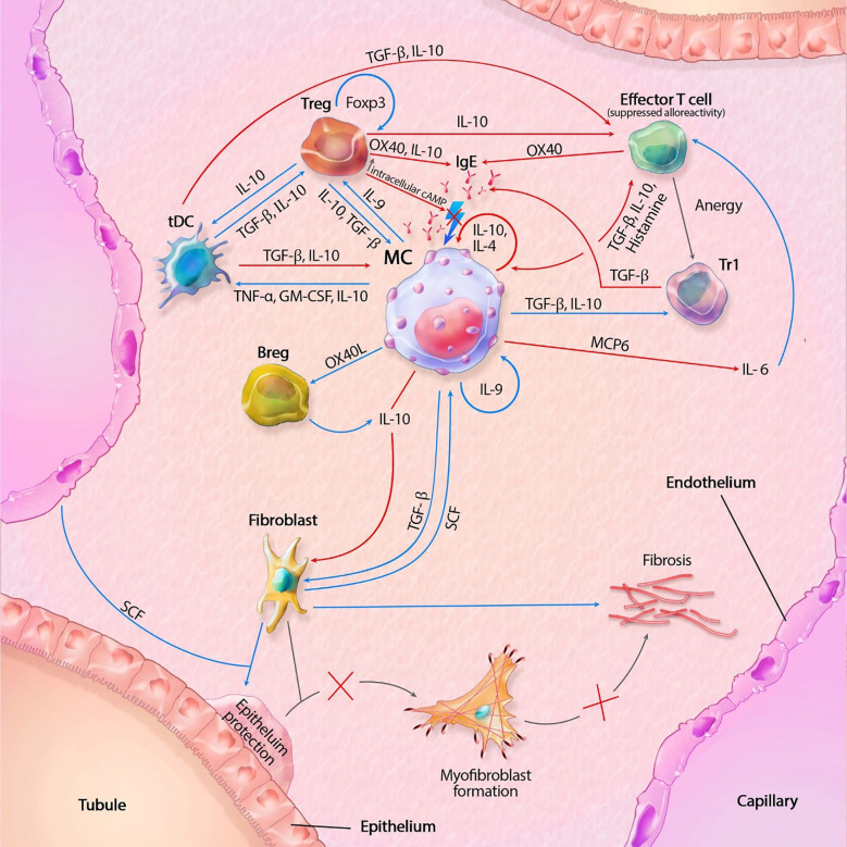
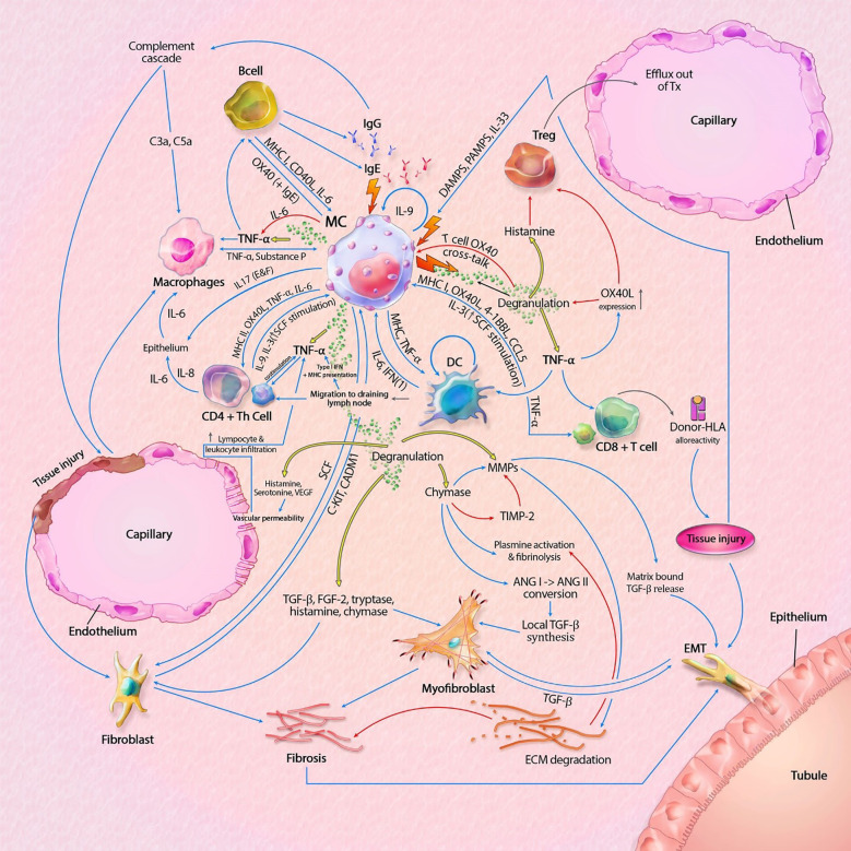
Mast cells (MCs) are innate immune cells with a versatile set of functionalities, enabling them to orchestrate immune responses in various ways. Aside from their known role in allergy, they also partake in both allograft tolerance and rejection through interaction with regulatory T cells, effector T cells, B cells and degranulation of cytokines and other mediators. MC mediators have both pro- and anti-inflammatory actions, but overall lean towards pro-fibrotic pathways. Paradoxically, they are also seen as having potential protective effects in tissue remodeling post-injury. This manuscript elaborates on current knowledge of the functional diversity of mast cells in kidney transplants, combining theory and practice into a MC model stipulating both protective and harmful capabilities in the kidney transplant setting.

Keywords: fibrosis; kidney transplant; mast cell (MC); rejection; tolerance.


Conflict of interest statement

The authors declare that the research was conducted in the absence of any commercial or financial relationships that could be construed as a potential conflict of interest.

Figures

Figure 1
Figure 1
Mast cell (MC) interactions within the transplant during tolerance. FcϵRI activity is inhibited by TGF-β, IL-10 and OX40 ligation. Tregs also inhibit degranulation by lowering intracellular Ca2+ levels through increased cAMP. IL-10 suppresses alloreactivity within CD4+ and CD8+ T cells and promote anergy and regulatory functions of CD4+ T cells. IL-10 mediated inhibition of fibroblasts also inhibit subsequent formation of myofibroblasts. IL-10 with co-stimulation of IL-4 decrease MC proliferation, while IL-9 increases proliferation. GM-CSF, granulocyte-macrophage colony-stimulating factor; IL, interleukin; MCP6, mat cell protease 6; SCF, stem cell factor; tDC, tolerogenic dendritic cell; TGF-β, tissue growth factor beta; TNF-α, tissue necrotic factor alpha; Tr1, regulatory T cell type 1 (induced); Treg, regulatory T cell (natural); Blue lines symbolize activating pathways, red lines inhibitory pathways, gray lines symbolize subsequent events. Lighting icons are used in the most profound activation patterns, which are inhibited in tolerogenic environments.
Figure 2
Figure 2
Mast cell (MC) interactions within the graft during rejection. Pathways can include both cytokines (like TNF-α) and membrane bound interaction (like MHC I-TLR interaction). MC-T cell interaction through OX40L-OX40 cross-linking inhibits MC degranulation, represented by the inhibitory pathway towards degranulation. Innate immune cells can also result in tissue injury, which is not shown in this model. Interaction between APCs, T cells and B cells, resulting in antigen production is also not shown in this model. The model shows almost no inhibitory pathways, explaining the progressive state of fibrosis within KTx even when immunosuppressive drugs are taken. Detailed description of the model can be found within the text. ANG, angiotensin; C3a/C5a, complement component; ECM, extracellular matrix; EMT, epithelial-mesenchymal transition; FGF-2; fibroblast growth factor-2; Ig, immunoglobulin; IL, interleukin; MHC, major histocompatibility complex; MMPs, matrix metalloproteinase; SCF, stem cell factor; tDC, tolerogenic dendritic cell; TGF-β, tissue growth factor beta; Th cell, T helper cell; TIMP-2, tissue inhibitor of metalloproteinase-2; TNF-α, tissue necrotic factor alpha; Treg, regulatory T cell (natural); VEGF, Vascular Endothelial Growth Factor. Blue lines symbolize activating pathways, red lines inhibitory pathways, yellow lines represent pre-formed mediators within MCs. Grey lines represent subsequent events. Lighting icons are used in the most profound activation patterns.

References

    1. Hart A, Lentine KL, Smith JM, Miller JM, Skeans MA, Prentice M, et al. OPTN/SRTR 2019 annual data report: Kidney. Am J Transplant (2021) 21 Suppl 2:21–137. doi: 10.1111/ajt.16502 - DOI - PubMed
    1. Nankivell BJ, Agrawal N, Sharma A, Taverniti A, P'Ng CH, Shingde M, et al. The clinical and pathological significance of borderline T cell-mediated rejection. Am J Transplant (2019) 19(5):1452–63. doi: 10.1111/ajt.15197 - DOI - PubMed
    1. Wekerle T, Segev D, Lechler R, Oberbauer R. Strategies for long-term preservation of kidney graft function. Lancet (2017) 389(10084):2152–62.doi: 10.1016/S0140-6736(17)31283-7 - DOI - PubMed
    1. Coemans M, Callemeyn J, Naesens M. Long-term survival after kidney transplantation. N Engl J Med (2022) 386(5):497–8. doi: 10.1056/NEJMc2115207sa1 - DOI - PubMed
    1. Hariharan S, Israni AK, Danovitch G. Long-term survival after kidney transplantation. N Engl J Med (2021) 385(8):729–43. doi: 10.1056/NEJMra2014530 - DOI - PubMed

Publication types