Review
. 2023 Sep 20;40(9):1521-1549.
doi: 10.1039/d2np00086e.

S-Adenosylmethionine: more than just a methyl donor

Affiliations
Review

S-Adenosylmethionine: more than just a methyl donor

Yu-Hsuan Lee et al. Nat Prod Rep. .

Abstract

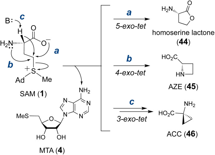
Covering: from 2000 up to the very early part of 2023S-Adenosyl-L-methionine (SAM) is a naturally occurring trialkyl sulfonium molecule that is typically associated with biological methyltransfer reactions. However, SAM is also known to donate methylene, aminocarboxypropyl, adenosyl and amino moieties during natural product biosynthetic reactions. The reaction scope is further expanded as SAM itself can be modified prior to the group transfer such that a SAM-derived carboxymethyl or aminopropyl moiety can also be transferred. Moreover, the sulfonium cation in SAM has itself been found to be critical for several other enzymatic transformations. Thus, while many SAM-dependent enzymes are characterized by a methyltransferase fold, not all of them are necessarily methyltransferases. Furthermore, other SAM-dependent enzymes do not possess such a structural feature suggesting diversification along different evolutionary lineages. Despite the biological versatility of SAM, it nevertheless parallels the chemistry of sulfonium compounds used in organic synthesis. The question thus becomes how enzymes catalyze distinct transformations via subtle differences in their active sites. This review summarizes recent advances in the discovery of novel SAM utilizing enzymes that rely on Lewis acid/base chemistry as opposed to radical mechanisms of catalysis. The examples are categorized based on the presence of a methyltransferase fold and the role played by SAM within the context of known sulfonium chemistry.


Conflict of interest statement

The authors declare no conflicts of interest.

Figures

Fig. 1
Fig. 1. Distinct uses of SAM in enzymatic reactions.
Fig. 2
Fig. 2. Summary of SAM-dependent alkylation reactions, showing the by-product generated following each group transfer. (A) Methyl transfer yields S-adenosyl homocysteine (SAH, 3), (B) ACP (3-amino-3-carboxypropyl) transfer yields 5′-methylthioladenosine (MTA, 4) and (C) adenosyl transfer yields l-methionine (5).
Fig. 3
Fig. 3. Aminopropyl group transfer during the biosynthesis of spermidine. SAM is first decarboxylated to form decarboxy-SAM (dc-SAM, 6) before the 3-aminopropyl group is transferred to putrescine to yield spermidine (7).
Fig. 4
Fig. 4. (A) Nicotianamine synthase (NAS) catalyzes the formation of nicotianamine (8) from three molecules of SAM. (B) Molecular mechanism proposed for MtNAS during the biosynthesis of thermonicotianamine (tNA, 9). (C) Crystallographic snapshot of MtNAS (PDB ID: 3FPF) complexed with MTA (pink) and tNA (tan). Nucleophilic displacement was facilitated by deprotonation of the substrate nitrogen by Tyr107 and Glu81 (cyan). (D) The NAS-like enzyme CntL catalyzes transfer of one ACP to d-His (13) providing x-NA (14) as the precursor of bacterial metallophore, staphylopine (12).
Fig. 5
Fig. 5. Aminopropylation during maturation of microcin C (15). MccD can catalyze transfer of an ACP group onto McC1120 (16), which can further be decarboxylated in a reaction catalyzed by MccE (route a). An alternative pathway starts with the formation of dc-SAM catalyzed by MccE followed by MccD mediated aminopropyl transfer (route b).
Fig. 6
Fig. 6. Other natural products with ACP groups derived from SAM.
Fig. 7
Fig. 7. Enzymatic functions of (A) Tsr3 and (B) its closest homolog Trm10. Tsr3 catalyzes ACP modification of a pseudouridine residue whereas Trm10 catalyzes methylation of a guanosine or adenosine residue. (C) Structure of wybutosine highlighting the ACP moiety transferred under the action of Tyw2. Solvent accessible area of (D) Tsr3 (PDB ID: 5APG) and (E) Trm10 (PDB ID: 4JWF) is shown colored with the most hydrophilic residues in cyan to the most hydrophobic in gold. In Tsr3, the ACP side chain of Se-SAM (pink) is exposed to solvent and stabilized by hydrophilic interactions. Asp70, Ser72 and Trp73 in the DTW domain are in purple. In contrast, the ACP side chain of SAH (pink) in Trm10 is buried, and the entrance to the binding cavity is lined with hydrophobic residues. (F) Enzymatic function of TapT. (G) Sequence alignment of Tsr3 and TapT at the DTW domain (gray) (Ec: Escherichia coli; Vp: Variovorax paradoxus; Cs: Clostridium saccharoperbutylacetonicum; Vd: Vulcanisaeta distributa; Ss: Sulfolobus solfataricus; Hs: Homo sapiens; Sc: Saccharomyces cerevisiae).
Fig. 8
Fig. 8. (A) Enzymatic functions of CmoA and CmoB. Two CmoA homologs TglE and MccSBam also catalyze the formation of 29. (B) Crystal structure of Cx-SAM bound to CmoB (PDB ID: 4QNV). The carboxymethyl moiety of Cx-SAM is coordinated by Tyr200, Lys91 and Arg315. The unique 100 residues at the N-terminus of CmoB are shown in green.
Fig. 9
Fig. 9. SAM-dependent carboxymethylation during the biosynthesis of (A) 3-thiaglutamate (31) and (B) McC-like peptidyl cytidylate (34).
Fig. 10
Fig. 10. (A) Adenosyl transfer reactions catalyzed by FlA, SalL and DUF proteins. The resulting 5′-deoxy-5′-haloadenosine (36, 37) is a precursor to halogen containing natural products (39–42). (B) In the active site of SalL-Y70T mutant (PDB ID: 2Q6O), SAM (1, pink) and chloride (green) are aligned and poised for SN2 reaction.
Fig. 11
Fig. 11. A similar overall structure was observed for three adenosyl transfer enzymes. (A) The product complex between fluorinase FlA/36/5 (PDB ID: 1RQR). (B) The product complex between chlorinase SalL/37/5 (PDB ID: 2Q6I). (C) The product complex of a DUF62 protein from Pyrococcus horikoshii OT3 with 38 (PDB ID: 1WU8). SAM-derived products (i.e., 5 and 36–38) have a gray surface and the conserved His-Arg-Asp triad in DUF62 proteins is colored cyan.
Fig. 12
Fig. 12. Intramolecular cyclization of the SAM aminocarboxypropyl moiety into homoserine lactone (44, via route a), azetidine 2-carboxylic acid (AZE, 45, via route b) and 1-aminocyclopropane-1-carboxylic acid (ACC, 46, via route c), each with elimination of MTA (4).
Fig. 13
Fig. 13. Examples of natural products that contain an azetidine 2-carboxylate (AZE) moiety.
Fig. 14
Fig. 14. The proposed two enzyme cascade for C10P and C10Q catalyzed formation of the cyclopropane ring during biosynthesis of CC-1065 (51) and related compounds (52 & 53).
Fig. 15
Fig. 15. Cation-induced cyclizations catalyzed by (A) canonical terpene cyclases and by (B) cyclopropane fatty acid synthases.
Fig. 16
Fig. 16. Mechanism proposed for the SodC catalyzed cyclization of FPP.
Fig. 17
Fig. 17. (A) Methylation induced cyclization catalyzed by TleD in teleocidin biosynthesis. Crystal structures of (B) TleD hexamer (PDB ID: 5GM2) and (C) TleD active site formed at the interface of two monomers (green & blue) with bound teleocidin A1 (72, tan) and product SAH (pink).
Fig. 18
Fig. 18. (A) Mechanism of Ecm18 catalyzed rearrangement of a disulfide bridge to a thioacetal. (B) Hydrophobic surface of the Ecm18 substrate binding pocket (PDB ID: 4NEC) colored according to most hydrophobic (gold) and most hydrophilic (cyan) residues. (C) Ecm18/76/SAH ternary complex. The distance between the sulfur of SAH and the methyl group in 76 is consistent with the proposed methyltransfer reaction. The basic nitrogen on His115 (cyan) is near the disulfide bridge and could facilitate ylide 80 formation via deprotonation of the methylated sulfonium intermediate 79.
Fig. 19
Fig. 19. (A) Methylation induced cyclization in pyrroloindoline alkaloid biosynthesis. (B) Synthesis of pyrroloindolines via alkylation–cyclization cascade.
Fig. 20
Fig. 20. (A) LepI catalyzed dehydration–cyclization cascade during leporin C biosynthesis. Although the stereochemistry at C7 in 91 is currently unclear, it has been shown that the diastereomer of 91 is not accepted by LepI. Comparison of crystal structures of (B) LepI/SAM/89 complex (PDB ID: 6IX9) and (C) OxaC/SFG/95 complex (PDB ID: 5W7S). In LepI, SAM is too far from the product (89, in tan) for methyl transfer; however, in the methyltransferase OxaC, the substrate meleagrin (95) (tan) is located in between SFG and the His313-Glu369 dyad. In contrast, the catalytic histidine is substituted with Arg295 in LepI such that deprotonation cannot occur. Green ribbons in LepI are residues from the second monomeric peptide. (D) OxaC catalyzed methylation during formation of oxaline (96). (E) Structure of sinefungin (94). (F) Cyclization of 98 catalyzed by FinI. (G) Sequence alignment of LepI, FinI and OxaC highlighting the conserved SAM binding arginine residue (Arg310) that interacts with SAM, the catalytic base (His313) in OxaC (in gray boxes) and the essential arginine (Arg295/Arg313) in the pericyclases in red.
Fig. 21
Fig. 21. Crystal structures of (A) LepI (PDB ID: 6IX5), (B) SpnF (PDB ID: 4PNE) and (C) SpnL (PDB ID: 7V6H). Each enzyme contains a C-terminal Rossmann fold catalytic domain containing seven β-strands (cyan) surrounded by five α-helices (blue), while the N-terminal domains differ between each protein. The cofactor, SAM (1) for LepI and SAH (3) for SpnF and SpnL (pink with a gray surface), binds similarly at the C-terminal domain of all three enzymes. The substrate analog (90) bound in the LepI active site is shown with a green surface.
Fig. 22
Fig. 22. Biosynthesis of spinosyn A highlighting the [4 + 2] cycloaddition catalyzed by SpnF and two proposed mechanisms for the transannulation catalyzed by SpnL.
Fig. 23
Fig. 23. (A) C13-fluorinated substrate (109) leads to covalent modification of SpnL. (B) Crystal structure of SpnL/SAH binary complex (PDB ID: 7V6H) highlighting the SAH (3) binding site and the catalytic Cys60 (carbons as cyan and sulfur as yellow spheres), which is buried in the protein interior.
Fig. 24
Fig. 24. (A) Cyclization catalyzed by IccD during the biosynthesis of ilicicolin H. (B) Dehydration–cyclization reactions catalyzed by SAM-independent pericyclases that each adopt a SAM-binding fold.
Fig. 25
Fig. 25. SlnM catalyzes spirocyclization during salinomycin biosynthesis. SAM is proposed to stabilize an anionic aspartate residue via electrostatic interactions to facilitate substrate protonation.
Fig. 26
Fig. 26. (A) Selected anthracycline natural products generated via RdmB or DnrK catalysis. (B) Proposed mechanism for RdmB/DnrK reaction with 127. (C) Activity of methyltransferase DnrK with 135.
Fig. 27
Fig. 27. (A) Comparison of the binding of SAM (light green)/131′ (green) in RdmB and SAH (salmon)/136 (pink) in DnrK. (B) Sequence alignment of DnrK and RdmB at the α-helix that contains the aromatic residue, highlighting the position of serine insertion in DnrK-Ser. The orientation of Phe296 in DnrK or Phe300 in RdmB dictates the catalytic activity being methylation or hydroxylation. (C) Clip view of the RdmB/SAM/131′ complex (PDB ID: 1XDS). (D) Clip view of the DnrK/SAH/136 complex (PDB ID: 1TW2). The channel to the bulk solvent is blocked in RdmB due to the presence of Phe300 (cyan), whereas it is opened in DnrK.
Fig. 28
Fig. 28. NDUFAF5 catalyzes modification of the substrate protein NDUFS7 at Arg73. The precise site of hydroxylation is unknown.
Fig. 29
Fig. 29. Proposed mechanism for BrvO catalyzed decarboxylation.
Fig. 30
Fig. 30. (A) Proposed mechanism for CmoA catalysis. (B) Clip view of Cx-SAM/CmoA complex structure (PDB ID: 4GEK) highlighting the inner cavity for Cx-SAM binding. Computational substrate docking models have suggested that SAM adopts a similar binding configuration as that of the product Cx-SAM. Prephenate (146) is predicted to bind in the hydrophobic pocket adjacent to SAM (green arrow). In the zoomed in region, the surface coloring indicates hydrophobicity (hydrophobic in gold and hydrophilic in cyan). The red arrow indicates the position of the methyl group of SAM, which is surrounded by a hydrophobic environment.
Fig. 31
Fig. 31. Proposed mechanism of QueA catalyzed ribose transfer reaction.
Fig. 32
Fig. 32. (A) Transamination of SAM catalyzed by BioA. (B) Sequence alignment of RquA with related functional methyltransferases. RquA has two essential aspartate residues (Asp118 and Asp143, numbers in blue) for SAM binding whereas the DXXXGXG (red box) motif typically found in Rossmann fold MTs is missing in RquA. (C and D) Two proposed mechanisms for RquA catalyzed transamination during the biosynthesis of rhodoquinone (159).
Fig. 33
Fig. 33. Proposed mechanism for the transformation of SAM to ACC (46) and Me-ACC (170) catalyzed by GnmY and Orf29/Orf30, respectively.
Fig. 34
Fig. 34. Natural products that contain SAM-derived cyclopropane moieties.
Fig. 35
Fig. 35. Natural products that contain the ADR-GlyU disaccharide. The bold alkyl chain indicates the putative methionine-derived aminopropyl moiety.
Fig. 36
Fig. 36. Proposed mechanism for Mur24 catalyzed reaction.
Fig. 37
Fig. 37. Proposed mechanism of SbzP catalyzed transformation of SAM (1) and β-NAD (187) into 192, which serves as the precursor to 6-azatetrahydroindane containing natural products (184–186).
Fig. 38
Fig. 38. Proposed mechanism of CqsA reaction.
Fig. 39
Fig. 39. Proposed mechanism for the ClbI catalyzed cyclopropylation during the formation of colibactin biosynthetic intermediate 203.
None
Yu-Hsuan Lee
None
Daan Ren
None
Byungsun Jeon
None
Hung-wen Liu

References

    1. Lu S. C. Int. J. Biochem. Cell Biol. 2000;32:391–395. doi: 10.1016/S1357-2725(99)00139-9. - DOI - PubMed
    1. Chiang P. K. Gordon R. K. Tal J. Zeng G. C. Doctor B. P. Pardhasaradhi K. McCann P. P. FASEB J. 1996;10:471–480. doi: 10.1096/fasebj.10.4.8647346. - DOI - PubMed
    1. Loenen W. A. M. Biochem. Soc. Trans. 2006;34:330–333. doi: 10.1042/BST0340330. - DOI - PubMed
    1. Liscombe D. K. Louie G. V. Noel J. P. Nat. Prod. Rep. 2012;29:1238–1250. doi: 10.1039/C2NP20029E. - DOI - PubMed
    1. Blum M. Chang H.-Y. Chuguransky S. Grego T. Kandasaamy S. Mitchell A. Nuka G. Paysan-Lafosse T. Qureshi M. Raj S. Richardson L. Salazar G. A. Williams L. Bork P. Bridge A. Gough J. Haft D. H. Letunic I. Marchler-Bauer A. Mi H. Natale D. A. Necci M. Orengo C. A. Pandurangan A. P. Rivoire C. Sigrist C. J. A. Sillitoe I. Thanki N. Thomas P. D. Tosatto S. C. E. Wu C. H. Bateman A. Finn R. D. Nucleic Acids Res. 2021;49:D344–D354. doi: 10.1093/nar/gkaa977. - DOI - PMC - PubMed

Publication types