. 2023 Jan 1;13(1):e2023018.
doi: 10.5826/dpc.1301a18.

Lead Time from First Suspicion of Malignant Melanoma in Primary Care to Diagnostic Excision: a Cohort Study Comparing Teledermatoscopy and Traditional Referral to a Dermatology Clinic at a Tertiary Hospital

Affiliations

Lead Time from First Suspicion of Malignant Melanoma in Primary Care to Diagnostic Excision: a Cohort Study Comparing Teledermatoscopy and Traditional Referral to a Dermatology Clinic at a Tertiary Hospital

Karina Schultz et al. Dermatol Pract Concept. .

Abstract

Introduction: The increasing use of teledermatoscopy in clinical practice has led to demands to evaluate the effects of this new technology on traditional healthcare systems.

Objectives: To study lead times from first consultation in primary care to diagnostic excision of suspected malignant melanoma lesions in traditional referrals to a tertiary hospital-based dermatology clinic compared with mobile teledermatoscopy referrals.

Methods: A retrospective cohort study design was used. Data on sex, age, pathology, caregivers, clinical diagnosis, date for first visit to primary care unit, and date for diagnostic excision were collected from medical records. Patients managed through traditional referral (n=53) were compared with patients managed at primary care units using teledermatoscopy (n=128) regarding lead time from first visit to diagnostic excision.

Results: Mean time from date of first visit at primary care unit to diagnostic excision did not differ between the traditional referral and teledermatoscopy groups (16.2 vs. 15.7 days, median 10 vs. 13 days, p=0.657). Lead times from date of referral to diagnostic excision did not significantly differ (15.7 vs. 12.8 days, median 10 vs. 9 days, p=0.464).

Conclusions: Our study indicates that lead time to diagnostic excision for patients with suspected malignant melanoma managed by teledermatoscopy was comparable and not inferior to that of the traditional referral pathway. If teledermatoscopy is used at first consultation in primary care, it could potentially be more efficient than traditional referral.


Conflict of interest statement

Competing Interests: None.

Figures

Figure 1
Figure 1
Process of mobile teledermatoscopy as used in the project. The primary care physician examines the patient, takes clinical and dermatoscopic images (A and B), and fills in clinical information which is then packaged together in the mobile app and sent encrypted to a database. Photo by Oscar Segerström, Medicinsk bild, Karolinska Hospital.
Figure 2
Figure 2
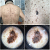
Photographs of a teledermatoscopy case. (A) An overview, (B) a close-up, and two dermatoscopic images ((C) polarized, (D) unpolarized) are collected and uploaded in the mobile application together with background information. Histopathologic diagnosis: Lentiginous malignant melanoma in situ described in pathology report as an extensive atypical junction melanocytic proliferation, lentiginous and nested, with frequent rete fusion, multifocal pagetoid upgrowth and early involvement of adnexal structures. The lesion focally blends with areas of seborrheic keratosis-like epidermal reaction and a small benign intradermal nevus is noted at the periphery of the main lesion.
Figure 3
Figure 3
Example of a teledermatoscopy case. The primary care physician fills in clinical information. Two dermatologists assess the case independently and provide a detailed description of the dermatoscopic findings, a provisional diagnosis, and a consensus recommendation for further diagnostic action.
Figure 4
Figure 4
Study flowchart. DU – dermatology unit, PC – primary care, PCU – primary care unit, SU – surgery unit, SC – standardized care, tertiary DU – Södersjukhuset dermatology unit. * SC pathway ends at face-to-face visit.

References

    1. Eriksson H, Nielsen K, Vassilaki I, et al. Trend Shifts in Age-Specific Incidence for In Situ and Invasive Cutaneous Melanoma in Sweden. Cancers (Basel) 2021;13(11) doi: 10.3390/cancers13112838. - DOI - PMC - PubMed
    1. Lyth J, Mikiver R, Nielsen K, Isaksson K, Ingvar C. Prognostic instrument for survival outcome in melanoma patients: based on data from the population-based Swedish Melanoma Register. Eur J Cancer. 2016;59:171–178. doi: 10.1016/j.ejca.2016.02.029. - DOI - PubMed
    1. Tensen E, van der Heijden JP, Jaspers MW, Witkamp L. Two Decades of Teledermatology: Current Status and Integration in National Healthcare Systems. Curr Dermatol Rep. 2016;5:96–104. doi: 10.1007/s13671-016-0136-7. - DOI - PMC - PubMed
    1. Lee KJ, Finnane A, Soyer HP. Recent trends in teledermatology and teledermoscopy. Dermatol Pract Concept. 2018;8(3):214–223. doi: 10.5826/dpc.0803a13. - DOI - PMC - PubMed
    1. Fernemark H, Skagerström J, Seing I, Ericsson C, Nilsen P. Digital consultations in Swedish primary health care: a qualitative study of physicians’ job control, demand and support. BMC Fam Pract. 2020;21(1):241. doi: 10.1186/s12875-020-01321-8. - DOI - PMC - PubMed

LinkOut - more resources