Review
. 2023 May-Jun;39(3):e91-e96.
doi: 10.1097/IOP.0000000000002347. Epub 2023 Mar 9.

Periorbital Pyoderma Gangrenosum Associated With a Cocaine-Induced Midline Destructive Lesion: Case Report and Review of the Literature

Affiliations
Review

Periorbital Pyoderma Gangrenosum Associated With a Cocaine-Induced Midline Destructive Lesion: Case Report and Review of the Literature

Marissa K Shoji et al. Ophthalmic Plast Reconstr Surg. 2023 May-Jun.

Abstract

A 72-year-old woman with a history of chronic cocaine use presented 9 months after a dog bite with a large facial ulceration and absent sinonasal structures. Biopsies were negative for infectious, vasculitic, or neoplastic pathologies. The patient was lost to follow up for 15 months and returned with a significantly larger lesion despite abstinence from cocaine. Additional inflammatory and infectious workup was negative. Intravenous steroids were administered with clinical improvement. Therefore, she was diagnosed with pyoderma gangrenosum and cocaine-induced midline destructive lesion due to cocaine/levamisole. Pyoderma gangrenosum is a rare dermatologic condition that uncommonly involves the eye and ocular adnexa. Diagnosis involves clinical examination, response to steroids, exclusion of infectious or autoimmune conditions, and identifying potential triggers including cocaine/levamisole. This report highlights a rare presentation of periorbital pyoderma gangrenosum causing cicatricial ectropion associated with concomitant cocaine-induced midline destructive lesion and reviews important aspects of clinical manifestations, diagnosis, and management of pyoderma gangrenosum and cocaine/levamisole autoimmune phenomenon.


Conflict of interest statement

Dr. Thaller reports royalties from Thieme and Springer book publishing companies. The other authors have no relevant financial conflicts of interest. The authors alone are responsible for the content and writing of the paper.

Figures
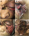
Figure 1.
Figure 1.
External examination of the patient on initial presentation demonstrates ulceration centrally with loss of the nasal bone, ethmoid sinus, septum, and proximal nasolacrimal system (A) with a large wound with undermined borders as well as central necrosis and crusting along the left temporal and infraorbital region resulting in a cicatricial ectropion (B). After being lost to follow-up for 15 months, the patient presented again with examination later demonstrating continued central ulceration (C) with progression of the cicatricial ectropion, and infraorbital and temporal wound with areas of necrosis and black eschar (D).
Figure 2.
Figure 2.
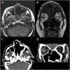
Magnetic resonance imaging axial (A) and coronal (B) views on initial presentation demonstrate absent sinonasal structures, full-thickness soft tissue and bony defects of the superior aspect of the external nose, and soft tissue enhancement and stranding in the inferomedial extraconal left orbit. Computerized tomography axial (C) and coronal (D) views of the patient fifteen months later after she was lost to follow-up continue to demonstrate absent sinonasal structures with soft tissue enhancement and premaxillary soft tissue thickening now extending into the left temporal scalp, preauricular region, and upper face.
Figure 3.
Figure 3.
Histopathology demonstrates a dermal lymphoplasmacytic cell inflammation with a neutrophilic inflammatory crust (A: hematoxylin and eosin, original magnification x200, B: hematoxylin and eosin, original magnification x600).
Figure 4.
Figure 4.
External examination of the patient after immunosuppression. Although she had initial progression of her disease due to being lost to follow-up and not using immunosuppressant medications, she was restarted on prednisone and cyclosporine and subsequently developed new pink granulation tissue covering her wound and improvement in the areas of eschar and necrosis.

References

    1. McElnea E, Stephenson K, Fulcher T. Pyoderma gangrenosum affecting the eye, orbit, and adnexa. A review. Orbit. 2018;37(1):26–31. doi:10.1080/01676830.2017.1353114 - DOI - PubMed
    1. Xu A, Balgobind A, Strunk A, Garg A, Alloo A. Prevalence estimates for pyoderma gangrenosum in the United States: An age- and sex-adjusted population analysis. J Am Acad Dermatol. 2020;83(2):425–429. doi:10.1016/j.jaad.2019.08.001 - DOI - PubMed
    1. Rose GE, Barnes EA, Uddin JM. Pyoderma gangrenosum of the ocular adnexa: A rare condition with characteristic clinical appearances. Ophthalmology. 2003;110(4):801–805. doi:10.1016/S0161-6420(02)01966-8 - DOI - PubMed
    1. Minocha R, Sebaratnam DF, Choi JY. Sweet’s syndrome following surgery: Cutaneous trauma as a possible aetiological co-factor in neutrophilic dermatoses. Australas J Dermatol. 2015;56(3):e74–e76. doi:10.1111/ajd.12142 - DOI - PubMed
    1. Maverakis E, Marzano AV, Le ST, et al. Pyoderma gangrenosum. Nat Rev Dis Primer. 2020;6(1):1–19. doi:10.1038/s41572-020-0213-x - DOI - PubMed