. 2023 Mar 14;120(11):e2219523120.
doi: 10.1073/pnas.2219523120. Epub 2023 Mar 9.

Divalent siRNAs are bioavailable in the lung and efficiently block SARS-CoV-2 infection

Affiliations

Divalent siRNAs are bioavailable in the lung and efficiently block SARS-CoV-2 infection

Vignesh N Hariharan et al. Proc Natl Acad Sci U S A. .

Abstract

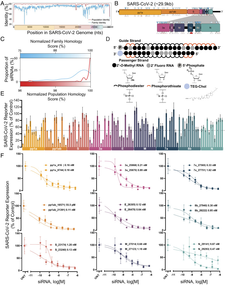
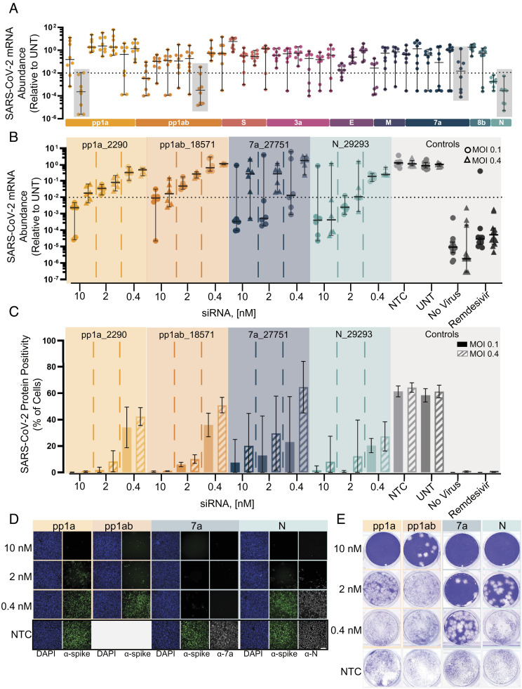
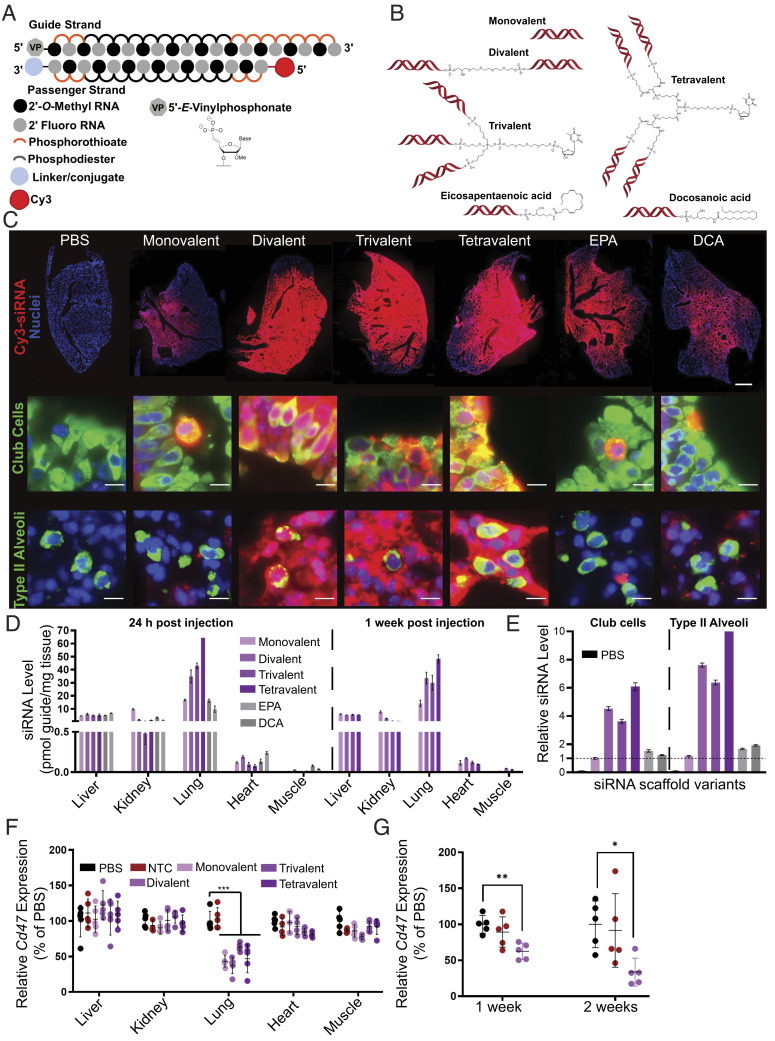
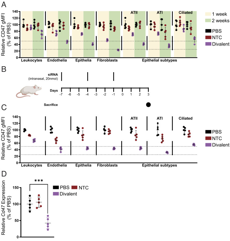
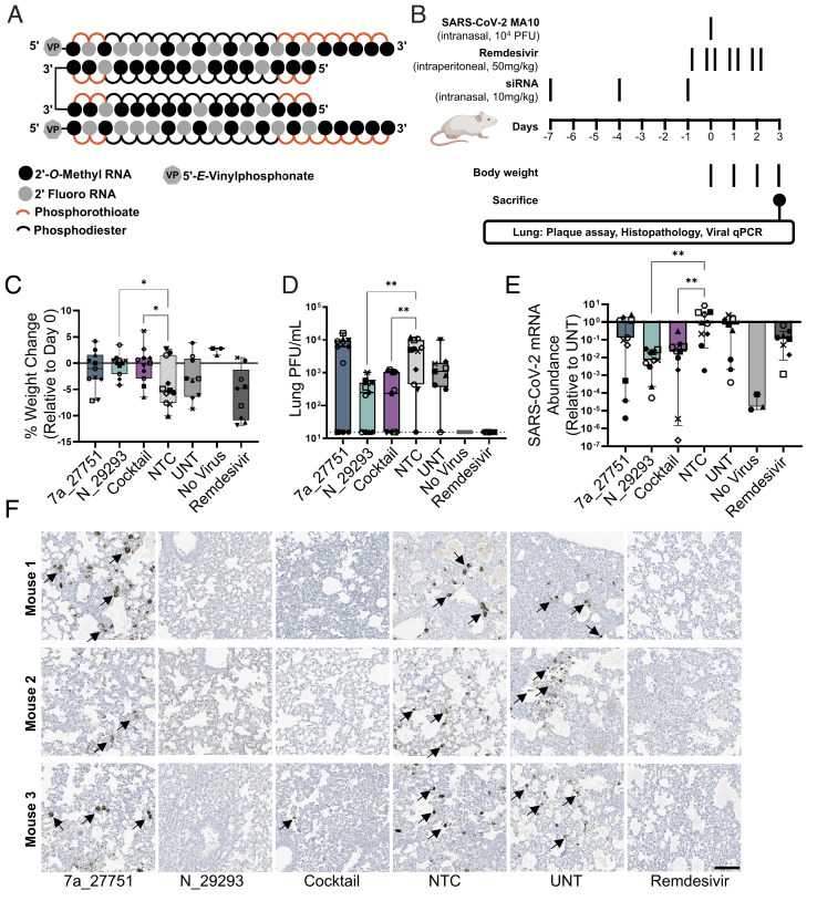
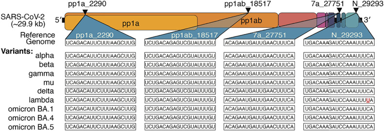
The continuous evolution of SARS-CoV-2 variants complicates efforts to combat the ongoing pandemic, underscoring the need for a dynamic platform for the rapid development of pan-viral variant therapeutics. Oligonucleotide therapeutics are enhancing the treatment of numerous diseases with unprecedented potency, duration of effect, and safety. Through the systematic screening of hundreds of oligonucleotide sequences, we identified fully chemically stabilized siRNAs and ASOs that target regions of the SARS-CoV-2 genome conserved in all variants of concern, including delta and omicron. We successively evaluated candidates in cellular reporter assays, followed by viral inhibition in cell culture, with eventual testing of leads for in vivo antiviral activity in the lung. Previous attempts to deliver therapeutic oligonucleotides to the lung have met with only modest success. Here, we report the development of a platform for identifying and generating potent, chemically modified multimeric siRNAs bioavailable in the lung after local intranasal and intratracheal delivery. The optimized divalent siRNAs showed robust antiviral activity in human cells and mouse models of SARS-CoV-2 infection and represent a new paradigm for antiviral therapeutic development for current and future pandemics.

Keywords: SARS-COV-2; antisense oligos; lung; mouse model; small interfering RNAs.


Conflict of interest statement

The authors have filed a patent application related to this work. G. Harold and Leila Y. Mathers Foundation (Mathers Foundation), UH | Harrington Discovery Institute, University Hospitals (HDI), HHS | NIH, HHS | NIH, HHS | NIH, Larry Ellison Foundation (LEF), Ono Pharmaceutical Foundation, Massachusetts Consortium on Pathogen Readiness (MCPR), U.S. Department of Defense (DOD), University of Massachusetts Chan Medical School, University of Massachusetts Chan Medical School.

Figures

Fig. 1.
Fig. 1.
Design and screening of fully chemically modified siRNAs targeting conserved SARS-CoV-2 gene regions. (A) Population identity indicates that from the alignment of the SARS-CoV-2 target genome with SARS-CoV-2 genomes from the COVID-19 patient. Family identity indicates that from the alignment of the SARS-CoV-2 target genome with the most closely related SARS-CoVs. All identities are averaged over a sliding window of fifty bases to facilitate visualization. (B) siRNAs were designed to target the open reading frame of the SARSCoV-2 genome indicated by triangles. Triangle colors correspond to the homology scores in (C)-upper triangles for family homology score and lower triangles for population homology score. (C) Each siRNA sequence was scored based on its homology to the SARS-CoV-2 population (red) and family (blue). (D) Schematic of the fully chemically modified siRNA. (E and F) Percent expression of SARS-CoV-2 reporter in HeLa cells 72 h after uptake of siRNA (n = 3; E: 1.5 µM, F: concentration indicated). Reporter expressions were assayed using the psiCHECK-2 reporter system for SARS-CoV-2. Data presented relative to UNT (mean ± SD of independent biological replicates). Target regions of SARS-CoV-2 are indicated in each graph. The dotted line indicates 50% silencing (E). IC50 values of each siRNA are indicated on each graph (F). UNT, untreated control.
Fig. 2.
Fig. 2.
Screening and lead characterization of fully chemically modified siRNAs in SARSCoV-2 infection model in human cells identify several lead candidates. (A) Relative abundance of viral mRNA in supernatant of A549ACE2/TMPRSS2 cells 48 h postinfection of SARS-CoV-2 with MOI 0.1. siRNAs were transfected 36 h prior to the infection (siRNA: n = 8, controls: n = 20; 10 nM). Abundance of viral mRNA was measured by qRT-PCR. Data presented relative to UNT (median ± 95% CI of independent biological replicates). Lead siRNAs selected in screening were further characterized (gray box). The dotted line indicates 99% reduction in mRNA abundance. (BE) Samples collected and analyzed from A549ACE2plus cells 48 h postinfection of SARS-CoV-2 with MOI 0.1 or 0.4. siRNAs were transfected 36 h prior to the infection (siRNA: n = 6, controls: n = 12; concentration indicated). Remdesivir with 5 µM was used as a positive control for antiviral activity. No virus represents cells which were not infected with virus. (B) Abundance of viral mRNA was measured by qRT-PCR. Data presented relative to UNT (median ± 95% CI of independent biological replicates). The dotted line indicates 99% reduction in mRNA abundance. (C) Percent of spike protein-positive cells was determined by immunofluorescence staining (mean ± SD of independent biological replicates). (D) Immunofluorescence images of the spike protein (α-spike; green), Orf7a protein (α-7a; gray), nucleocapsid protein (α-N; gray), and DAPI (blue). Original magnification, ×10. (Scale bar: 100 μm.) (E) Image of the viral plaque-forming assay conducted with supernatant. (Scale bar: 100 μm.) MOI, multiplicity of infection; NTC, nontargeting control; UNT, untreated control.
Fig. 3.
Fig. 3.
Local administration of multivalent fully chemically modified siRNA supports robust lung accumulation and safe and efficacious target gene silencing. (A) Schematic of the fully chemically modified siRNA. Multivalent linker or hydrophobic moiety is conjugated to the 3′ end of the passenger strand. Cy3 dye labeled to the 5′ end of the passenger strand. (B) Schematic of multivalent and hydrophobic siRNA scaffold. (C) Representative fluorescence images of lung after intratracheal injection of siRNA in mice sacrificed at 24 h postinjection (n = 2 mice; 3.75 mg/kg). (Top) Distribution of siRNA (red) with staining of DAPI (blue). Original magnification, ×5. (Scale bar: 1 mm.) (Middle and Bottom) Distribution of siRNA (red) with staining of club cells (green), type II alveoli (green), and DAPI (blue). Original magnification, ×40. (Scale bar: 10 µm.) Images collected at the same laser intensity and acquisition time. d, Tissue siRNA levels after intratracheal injection of siRNA in mice sacrificed at 24 h (Left) and 1 wk (Right) postinjection measured by PNA hybridization assay (n=2 mice; 3.75 mg/kg; mean ± SD of independent biological replicates). (E) siRNA uptake in club cells and type II alveoli after intratracheal injection of siRNA in mice sacrificed at 24 h quantified from microscope image (2 mice; 3.75 mg/kg). Bar colors same as D, with PBS group also shown in black on the left of each set. Data presented relative to monovalent siRNA of club cells (mean ± SEM of individual cells, n = 501 (PBS); 622 (monovalent); 798 (divalent); 946 (trivalent); 325 (tetravalent); 742 (EPA); and 545 (DCA) for club cells, n = 137 (PBS); 598 (monovalent); 753 (divalent); 796 (trivalent); 325 (tetravalent); 503 (EPA); and 326 (DCA) for type II alveoli). (F) Percent of Cd47 mRNA levels after intratracheal injection of siRNA targeting Cd47 in mice sacrificed at 1 wk postinjection (n = 5 mice; 7.5 mg/kg). mRNA levels were measured using QuantiGene assay, normalized to a housekeeping gene, Hprt, and presented relative to PBS (mean ± SD of independent biological replicates). (G) Percent of Cd47 mRNA level in bulk lung tissue after intratracheal injection of siRNA targeting Cd47 in mice sacrificed at 1 wk and 2 wk postinjection (3.25 mg/kg, n = 5 mice). mRNA levels were measured using QuantiGene assay, normalized to Hprt, and presented relative to PBS (mean ± SD of independent biological replicates).
Fig. 4.
Fig. 4.
Local administration of divalent fully chemically modified siRNA elicits potent gene silencing in multiple lung cell subtypes following intratracheal or intranasal injection. (A) Percentage CD47 geometric mean fluorescence intensity presented relative to PBS in leukocytes (CD45+), endothelial cells (CD45–CD31+CD326–), fibroblasts (CD45–CD31–CD326–CD140a+), type II alveoli (CD45–CD31–CD326medMHCII+), type I alveoli (CD45–CD31–CD326lowMHCII–), and ciliated cells (CD45–CD31–CD326hiMHCII–) after a single intratracheal injection of siRNA of 7.5 mg/kg sacrificed at 1 wk and 2 wk followed by flow cytometry analysis (n = 5 mice). (B) Schematic of intranasal multidose regimen optimized for expedited silencing. (C) Percentage CD47 geometric mean fluorescence intensity presented relative to PBS in leukocytes (CD45+), endothelial cells (CD45– D31+CD326–), fibroblasts (CD45–CD31–CD326–CD140a+), type II alveoli (CD45–CD31–CD326medMHCII+), type I alveoli (CD45–CD31–CD326lowMHCII–), and ciliated cells (CD45–CD31–CD326hiMHCII–) after intranasal injection of siRNA (20 nmol or ~10 mg/kg ×3) sacrificed at day 7 post first injection followed by flow cytometry analysis (n = 5 mice; ~10 mg/kg ×3). (D) Percent of Cd47 mRNA levels after intranasal injection of siRNA (20 nmol or ~10 mg/kg ×3) sacrificed at day 7 post first injection (n = 5 mice). mRNA levels were measured using QuantiGene assay, normalized to a housekeeping gene, Hprt, and presented relative to PBS (mean ± SD of independent biological replicates).
Fig. 5.
Fig. 5.
Intranasal administration of divalent siRNA is protective in a mouse model of SARS-CoV-2 infection. (A) Schematic of the fully chemically modified divalent siRNA. (B) Study design describing a mouse model of SARS-CoV-2 infection. Divalent siRNAs, 7a_27751, N_29293, and cocktail (1:1 mixture of 7a_27751 and N_29293), were given as pretreatment at days 7, 4, and 1 before viral infection with the SARS-CoV-2 strain MA10 (day 0) by intranasal administration (10 mg/kg/injection). Remdesivir was administered twice a day from day -1 to day 2 by intraperitoneal injection (50 mg/kg/injection) as a positive control. After viral infection, body weight was measured every day, and mice were sacrificed on day 3 (siRNA: n = 11, UNT: n = 9, no virus: n = 3, remdesivir: n = 9 mice). (C) Percent weight change on day 3 relative to day 0 (box plot: center line, median; box limits, upper and lower quartiles; whiskers, min to max; points, independent biological replicates). (D) Viral load in lung quantified by plaque-forming assay (box plot: center line, median; box limits, upper and lower quartiles; whiskers, min to max; points, independent biological replicates). The dotted line indicates the limit of detection (15.6 PFU/mL). (E) Abundance of viral mRNA in lung was measured by qRT-PCR, normalized to a housekeeping gene, Hprt. Data presented relative to UNT (median ± 95% CI of independent biological replicates). (F) Representative immunostaining images of mice lungs. Arrows indicate positive staining of nucleocapsid protein of SARS-CoV-2. Original magnification, ×20. (Scale bar: 100 µm.) CE, Individual mice are identified by a unique symbol. One-way ANOVA with Dunnett test for multiple comparisons (**P < 0.01, *P < 0.05). MOI, multiplicity of infection; NTC, nontargeting control; UNT, untreated control; PFU, plaque-forming units.
Fig. 6.
Fig. 6.
Lead siRNA compounds target all current SARS-CoV-2 variants of clinical concern. Triangles indicate target location in the coding regions of the SARS-CoV-2 genome. Sequences targeted by lead siRNA compounds and comparison with current SARS-CoV-2 variants of clinical concern. The mismatched base is denoted in red and underlined.

References

    1. Hadj Hassine I., Covid-19 vaccines and variants of concern: A review. Rev. Med. Virol. 32, e2313 (2021), 10.1002/rmv.2313. - DOI - PMC - PubMed
    1. Chakraborty C., Bhattacharya M., Sharma A. R., Present variants of concern and variants of interest of severe acute respiratory syndrome coronavirus 2: Their significant mutations in S-glycoprotein, infectivity, re-infectivity, immune escape and vaccines activity. Rev. Med. Virol. 32, e2270.
    1. Lauring A. S., Hodcroft E. B., Genetic variants of SARS-CoV-2-what do they mean? JAMA 325, 529–531 (2021). - PubMed
    1. Chakraborty C., et al. , Mode of transmission, and mutational landscape of newly emerging SARS-CoV-2 variants. mBio 12, e0114021 (2021). - PMC - PubMed
    1. Tao K., et al. , The biological and clinical significance of emerging SARS-CoV-2 variants. Nat. Rev. Genet 22, 757–773 (2021). - PMC - PubMed

Publication types

Supplementary concepts